
Bassett Collection of Stereoscopic Images of Human Anatomy
Dorsal aspect of right forearm
Relations of extensor muscles to ulnar border of forearm
Image #106-2
KEYWORDS: Forearm, Muscles and tendons, Vasculature, Overview.
Creative Commons
Stanford holds the copyright to the David L. Bassett anatomical images and has assigned Creative Commons license Attribution-Share Alike 4.0 International to all of the images.

For additional information regarding use and permissions, please contact the Medical History Center.
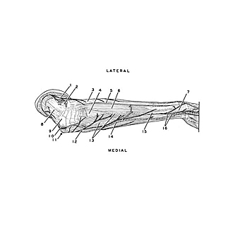
Dorsal aspect of right forearm
Relations of extensor muscles to ulnar border of forearm
The specimen shown in the previous view has been turned medially so that the relation of the ulna (15) to the extensor muscles (above) and the flexor muscles (below) is visible.
- Dorsal antebrachial cutaneous nerve
- Lateral epicondyle of humerus
- Common extensor digitorum muscle
- Anconeus muscle
- Extensor digiti minimi muscle
- Extensor carpi ulnaris muscle
- Styloid process of ulna
- Triceps brachii muscle
- Olecranon
- Ulnar nerve (in sulcus of ulnar nerve)
- Medial epicondyle of humerus
- Middle antibrachial cutaneous nerve
- Flexor carpi ulnaris muscle (ulnar head)
- Basilic vein
- Body of ulna
- Dorsal hand branch of ulnar nerve