Bassett Collection of Stereoscopic Images of Human Anatomy
Volar aspect of right hand
Common synovial sheath of flexor tendons (ulnar bursa)
Image #103-2
KEYWORDS: Fascia ligaments and tendons, Hand and fingers, Neuralnetwork, Peripheral nervous system, Vasculature.
Creative Commons
Stanford holds the copyright to the David L. Bassett anatomical images and has assigned Creative Commons license Attribution-Share Alike 4.0 International to all of the images.

For additional information regarding use and permissions, please contact the Medical History Center.
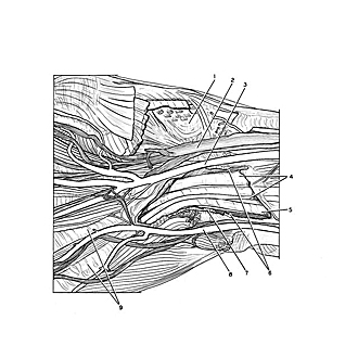
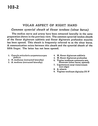
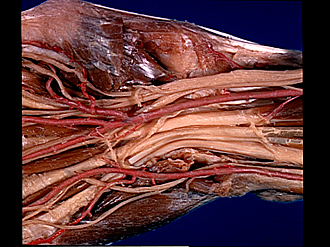
Volar aspect of right hand
Common synovial sheath of flexor tendons (ulnar bursa)
The median nerve and artery have been retracted laterally in the same preparation shown in the previous view. The common synovial tendon sheath of the flexor digitorum sublimis and flexor digitorum profundus muscles has been opened. This sheath is frequently referred to as the ulnar bursa. A communication exists between this sheath and the synovial sheath of the fifth finger. The latter has not been opened.
- Metacarpotrapezial joint capsule
- Median nerve (retracted laterally)
- Median artery (retracted laterally)
- Flexor digitorum superficialis
- Flexor digitorum profundus muscle
- Sheath of common tendon of flexor muscles (ulnar bursa, opened)
- Transverse carpal ligament (cut edge)
- Ulnar artery
- Tendon digital sheaths IV-V