
Bassett Collection of Stereoscopic Images of Human Anatomy
Volar aspect of right hand
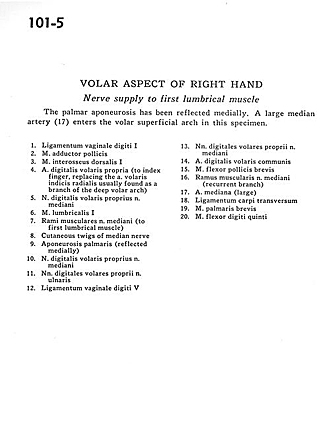
Nerve supply to first lumbrical muscle
Image #101-5
KEYWORDS: Hand and fingers, Neuralnetwork, Peripheral nervous system, Vasculature.
Creative Commons
Stanford holds the copyright to the David L. Bassett anatomical images and has assigned Creative Commons license Attribution-Share Alike 4.0 International to all of the images.

For additional information regarding use and permissions, please contact the Medical History Center.

Volar aspect of right hand
Nerve supply to first lumbrical muscle
The palmar aponeurosis has been reflected medially. A large median artery (17) enters the volar superficial arch in this specimen.
- Ligament of digital sheath I
- Adductor pollicis muscle
- Dorsal interosseous muscle I
- Proper palmar digital artery (to index finger, replacing the anterior radial index artery usually found as a branch of the deep anterior arch)
- Proper palmar digital nerve of median nerve
- Lumbrical muscle I
- Muscular branches of median nerve (to first lumbrical muscle)
- Cutaneous twigs of median nerve
- Palmar aponeurosis (reflected medially)
- Proper palmar digital nerve of median nerve
- Proper palmar digital nerves of ulnar nerve
- Ligament of digital sheath V
- Proper palmar digital nerves of median nerve
- Anterior common digital artery
- Flexor pollicis brevis muscle
- Muscular branch of median nerve (recurrent branch)
- Median artery (large)
- Transverse carpal ligament
- Palmaris brevis muscle
- Flexor digiti minimi muscle