Bassett Collection of Stereoscopic Images of Human Anatomy
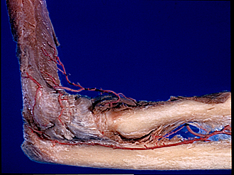
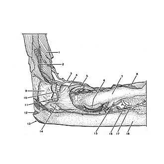
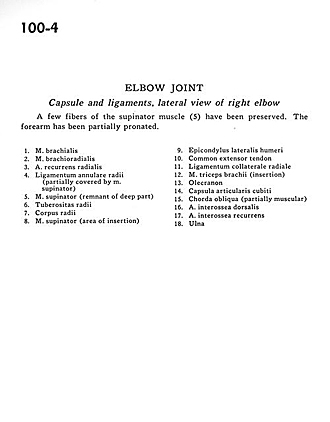
Elbow joint
Capsule and ligaments, lateral view of right elbow
Image #100-4
KEYWORDS: Elbow, Fascia ligaments and tendons, Muscles and tendons, Vasculature.
Creative Commons
Stanford holds the copyright to the David L. Bassett anatomical images and has assigned Creative Commons license Attribution-Share Alike 4.0 International to all of the images.

For additional information regarding use and permissions, please contact the Medical History Center.
Elbow joint
Capsule and ligaments, lateral view of right elbow
A few fibres of the supinator muscle (5) have been preserved. The forearm has been partially pronated.
- Brachialis muscle
- Brachioradialis muscle
- Recurrent radial artery
- Annular ligament of radius (partially covered by supinator muscle)
- Supinator muscle (remnant of deep part)
- Tuberosity of radius
- Body of radius
- Supinator muscle (area of insertion)
- Lateral epicondyle of humerus
- Common extensor tendon
- Radial collateral ligament
- Triceps brachii muscle (insertion)
- Olecranon
- Cubital joint capsule
- Oblique cord (partially muscular)
- Dorsal interosseous artery
- Recurrent interosseous artery
- Ulna