Bassett Collection of Stereoscopic Images of Human Anatomy
Exploration of the brain from its superior aspect
Interventricular foramina viewed from in front
Image #10-6
KEYWORDS:
Creative Commons
Stanford holds the copyright to the David L. Bassett anatomical images and has assigned Creative Commons license Attribution-Share Alike 4.0 International to all of the images.

For additional information regarding use and permissions, please contact the Medical History Center.

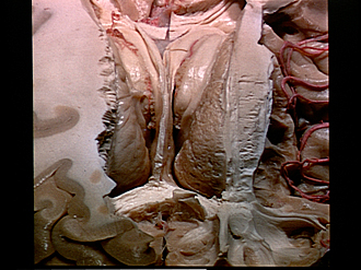
Exploration of the brain from its superior aspect
Interventricular foramina viewed from in front
The specimen is turned so that the direction of view is from the frontal pole. The position and relations of the interventricular foramina (of Monro) are demonstrated. Note
- Hippocampal commissure (cut through in midline)
- Crura of fornix
- Laminae affixae
- Corona radiata (partially dissected)
- Head of caudate nucleus
- Anterior horn lateral ventricle
- Genu corpus callosum (dissected parallel to fibers)
- Superior frontal gyrus (cut in horizontal plane)
- Anterior cerebral arteries lying within longitudinal fissure
- Caudate nucleus (tail)
- Anterior tubercle of thalamus
- Remnants of superior longitudinal fasciculus
- Interventricular foramen left (of Monro)
- Septum pellucidum (cut along line of attachment to corpus callosum)
- Superior occipitofrontal fasciculus
- Cingulum (cut across)
- Medial longitudinal stria right (cut off)