Bassett Collection of Stereoscopic Images of Human Anatomy
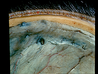
Exploration of the meninges and brain in situ
Close-up view of layers of scalp, parietal bone, dura mater and arachnoid granulations
Image #1-2
KEYWORDS: Bones cartilage joints, Brain, Meninges, Scalp.
Creative Commons
Stanford holds the copyright to the David L. Bassett anatomical images and has assigned Creative Commons license Attribution-Share Alike 4.0 International to all of the images.

For additional information regarding use and permissions, please contact the Medical History Center.

Exploration of the meninges and brain in situ
Close-up view of layers of scalp, parietal bone, dura mater and arachnoid granulations
- Epidermis
- Granular pit
- Arachnoid granulations
- Subaponeurotic space
- Venous lacuna (beneath dura mater)
- Branches Middle meningeal artery
- Dura mater
- Skin
- Radix (above pointer) and bulbus pili (below pointer)
- Superficial fascia (note numerous arteries and veins in this layer)
- Galea aponeurotica (firmly attached to Superficial fascia)
- Pericranium
- External plate of occipital bone
- Diploë
- Internal plate of occipital bone
- Emissary vessel of parietal bone