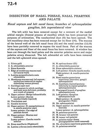
| 1
.
| Crista galli |
| 2
.
| Anterior ethmoidal artery |
| 3
.
| Frontal sinus |
| 4
.
| Edge of nasal mucosal membrane (of lateral wall) |
| 5
.
| Perpendicular lamina of ethmoid bone |
| 6
.
| Nasal mucosal membrane (of septum; epithelium removed to expose underlying venous plexus) |
| 7
.
| Nasal septal cartilage |
| 8
.
| Area of septum in which cartilage and bone were deficient, probably as a result of surgery |
| 9
.
| Upper pointer: Infraorbital margo Lower pointer: Nasolacrimal duct (opened) |
| 10
.
| Nasal vestibule |
| 11
.
| Incisive canal |
| 12
.
| Upper pointer: Nasopalatine nerve and posterior septal nasal artery Lower pointer: Cut edge of palatum durum |
| 13
.
| Upper pointer: Communicating nasal meatus Lower pointer: Inferior nasal concha |
| 14
.
| Maxillary sinus [Highmore] |
| 15
.
| Cribrosal lamina of ethmoid bone |
| 16
.
| Right optical nerve (II) |
| 17
.
| Posterior ethmoid artery |
| 18
.
| Left sphenoidal sinus |
| 19
.
| Left pointer: Medial nasal concha Right pointer: Posterior nasal septal artery |
| 20
.
| Rami nasales posteriores superiores [laterales] ganglii sphenopalatini |
| 21
.
| Maxillary nerve (turned postero-inferiorly to expose sphenopalatine ganglion) |
| 22
.
| Pharyngeal branch of sphenopalatine ganglion |
| 23
.
| Left pointer: Sphenopalatine ganglion Right pointer: Pterygopalatine canal nerve [Vidius] |
| 24
.
| Sphenopalatine nerves |
| 25
.
| Upper pointer: Posterior lateral nasal artery Lower pointer: Sphenopalatine artery |
| 26
.
| Posterior palatine nerve |
| 27
.
| Palatine levator veli muscle |
| 28
.
| Palatine tensor veli muscle |
| 29
.
| Major palatine artery (bony pterygopalatine canal removed) |
| 30
.
| Posterior superior alveolar nerves |
| 31
.
| Anterior palatine nerve |
| *
.
| [Legend above restored translation from Latin] |