Bassett Collection of Stereoscopic Images of Human Anatomy
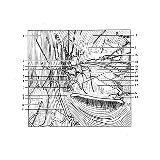
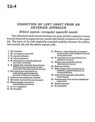
Dissection of left orbit from an anterior approach
Orbital septum; corrugator supercilii muscle
Image #52-4
KEYWORDS: Connective tissue, Eye, Face, Muscles and tendons, Peripheral nervous system, Vasculature.
Creative Commons
Stanford holds the copyright to the David L. Bassett anatomical images and has assigned Creative Commons license Attribution-Share Alike 4.0 International to all of the images.

For additional information regarding use and permissions, please contact Dr. Drew Bourn at dbourn@stanford.edu.

|
| ||||||||||||||||||||||||||||||||||||||||||||||
|
|