Bassett Collection of Stereoscopic Images of Human Anatomy
Osteology
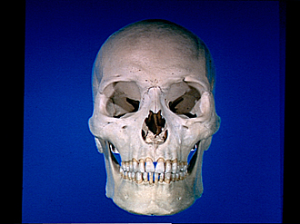
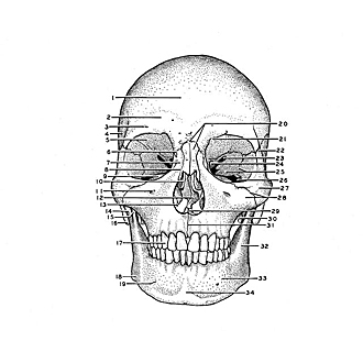
Skull, anterior view
Image #35-4
KEYWORDS: Bones cartilage joints, Cheek, Eye, Face, Mouth, Nose, Overview.
Creative Commons
Stanford holds the copyright to the David L. Bassett anatomical images and has assigned Creative Commons license Attribution-Share Alike 4.0 International to all of the images.

For additional information regarding use and permissions, please contact Dr. Drew Bourn at dbourn@stanford.edu.
|
| ||||||||||||||||||||||||||||||||||||||||||||||||||||||||||||||||||||||||

|
|