Bassett Collection of Stereoscopic Images of Human Anatomy
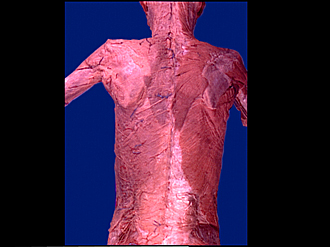
Dissection of thoracic and lumbosacral regions of back from a posterior approach
Superficial structures and external layer of muscles of back, general view
Image #212-1
KEYWORDS: Lumbar region, Muscles and tendons, Peripheral nervous system, Sacral region, Thoracic region, Vasculature, Vertebral column.
Creative Commons
Stanford holds the copyright to the David L. Bassett anatomical images and has assigned Creative Commons license Attribution-Share Alike 4.0 International to all of the images.

For additional information regarding use and permissions, please contact Dr. Drew Bourn at dbourn@stanford.edu.
|

| ||||||||||||||||||||||||||||||||||||||||

|
|