Bassett Collection of Stereoscopic Images of Human Anatomy
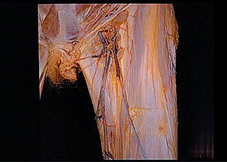
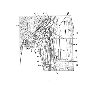
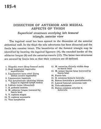
Dissection of anterior and medial aspects of thigh
Superficial structures overlying left femoral triangle, anterior view
Image #185-4
KEYWORDS: Fascia, Muscles and tendons, Thigh.
Creative Commons
Stanford holds the copyright to the David L. Bassett anatomical images and has assigned Creative Commons license Attribution-Share Alike 4.0 International to all of the images.

For additional information regarding use and permissions, please contact Dr. Drew Bourn at dbourn@stanford.edu.
|
| ||||||||||||||||||||||||||||||||||||||||||
|
|