Bassett Collection of Stereoscopic Images of Human Anatomy
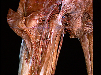
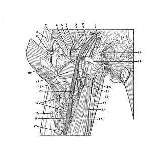
Dissection of posterior aspect of left thigh
Relations of sciatic nerve in upper part of thigh (continued)
Image #183-4
KEYWORDS: Peripheral nervous system, Thigh.
Creative Commons
Stanford holds the copyright to the David L. Bassett anatomical images and has assigned Creative Commons license Attribution-Share Alike 4.0 International to all of the images.

For additional information regarding use and permissions, please contact Dr. Drew Bourn at dbourn@stanford.edu.
|
| ||||||||||||||||||||||||||||||||||||||||||||||||||||||

|
|