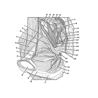
| 1
.
| Iliac branch of iliolumbar artery |
| 2
.
| Iliac crest |
| 3
.
| Obturator internus muscle |
| 4
.
| Obturator nerve |
| 5
.
| Iliopsoas muscle |
| 6
.
| Obturator artery |
| 7
.
| Superior pubic ramus |
| 8
.
| Pubic symphysis (sectioned) |
| 9
.
| Pubic arcuate ligament |
| 10
.
| Inferior pubic ramus |
| 11
.
| Ramus of ischium |
| 12
.
| Ramus communicans (to sacral nerve I) |
| 13
.
| Lumbosacral trunk |
| 14
.
| Sacral nerve I (ventral ramus) |
| 15
.
| Sympathetic trunk (pointer on ganglion) |
| 16
.
| Anterior (pelvic) sacral foramen (pointer indicates opening within sacral canal exposed by cutting through sacrum) |
| 17
.
| Rami communicantes |
| 18
.
| Left pointer: Sacral nerve II Right pointer: Sacral nerve III |
| 19
.
| Sacrum |
| 20
.
| Piriform muscle |
| 21
.
| Inferior gluteal artery |
| 22
.
| Sacral nerve IV |
| 23
.
| Upper pointer: Inferior rectal nerve Lower pointer: Pudendal nerve |
| 24
.
| Upper pointer: Branch of sacral nerve III to posterior femoral cutaneous nerve Lower pointer: Sciatic nerve |
| 25
.
| Nerve to obturator internus muscle (indicated in two places) |
| 26
.
| Coccygeus muscle |
| 27
.
| Location of ischial spine |
| 28
.
| Coccyx |
| 29
.
| Internal pudendal artery |
| 30
.
| Sacrotuberous ligament (cut off at attachment) |
| 31
.
| Ischial tuberosity |