Bassett Collection of Stereoscopic Images of Human Anatomy
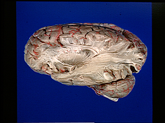
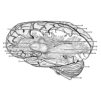
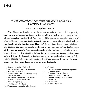
Exploration of the brain from its lateral aspect
External sagittal stratum
Image #14-2
KEYWORDS: Brain, Frontal lobe, Occipital lobe, Parietal lobe, Telencephalon, Temporal lobe.
Creative Commons
Stanford holds the copyright to the David L. Bassett anatomical images and has assigned Creative Commons license Attribution-Share Alike 4.0 International to all of the images.

For additional information regarding use and permissions, please contact Dr. Drew Bourn at dbourn@stanford.edu.
|
| ||||||||||||||||||||||||||||||||||||
|
|