Bassett Collection of Stereoscopic Images of Human Anatomy
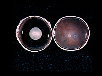
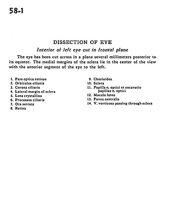
Dissection of eye
Interior of left eye cut in frontal plane
Image #58-1
KEYWORDS: Eye, Face.
Creative Commons
Stanford holds the copyright to the David L. Bassett anatomical images and has assigned Creative Commons license Attribution-Share Alike 4.0 International to all of the images.

For additional information regarding use and permissions, please contact the Medical History Center.
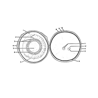
Dissection of eye
Interior of left eye cut in frontal plane
The eye has been cut across in a plane several millimeters posterior to its equator. The medial margins of the sclera lie in the center of the view with the anterior segment of the eye to the left.
- Optic (visual) part of retina
- Orbiculus ciliaris
- Corona ciliaris
- Lateral margin of sclera
- Crystalline lens
- Ciliary process
- Ora serrata
- Retina
- Choroid
- Sclera
- Papilla of optic nerve and hollowed area for papilla
- Macula lutea
- Fovea centralis
- Vorticose vein passing through sclera