
Bassett Collection of Stereoscopic Images of Human Anatomy
Joints of left ankle and foot
Metatarsophalangeal and interphalangeal joints, viewed from above
Image #204-3
KEYWORDS: Ankle, Bones joints cartilage, Foot and toes.
Creative Commons
Stanford holds the copyright to the David L. Bassett anatomical images and has assigned Creative Commons license Attribution-Share Alike 4.0 International to all of the images.

For additional information regarding use and permissions, please contact the Medical History Center.

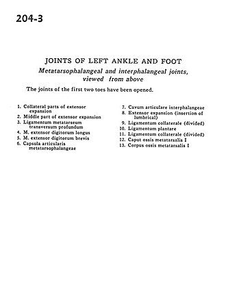
Joints of left ankle and foot
Metatarsophalangeal and interphalangeal joints, viewed from above
The joints of the first two toes have been opened.
- Collateral parts of extensor expansion
- Middle part of extensor expansion
- Deep transverse metatarsal ligament
- Extensor digitorum longus muscle
- Extensor digitorum brevis muscle
- Metatarsophalanx articular capsule
- Interphalangeal articular space
- Extensor expansion (insertion of lumbrical)
- Collateral ligament (divided)
- Plantar ligament
- Collateral ligament (divided)
- Head of 1st metatarsal bone
- Body of 1st metatarsal bone