
Bassett Collection of Stereoscopic Images of Human Anatomy
Dissection of anterior and medial aspects of thigh
Quadriceps femoris muscle.
Image #188-6
KEYWORDS: Muscles and tendons, Thigh.
Creative Commons
Stanford holds the copyright to the David L. Bassett anatomical images and has assigned Creative Commons license Attribution-Share Alike 4.0 International to all of the images.

For additional information regarding use and permissions, please contact the Medical History Center.
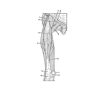

Dissection of anterior and medial aspects of thigh
Quadriceps femoris muscle.
The components of the right quadriceps femoris have been separated from each other to show their relations in a specimen from which most of the other muscles of the thigh have been removed. In the following views of this sequence the major features of this muscle complex are illustrated.
- Gluteus medius muscle
- Iliopsoas muscle
- Rectus femoris muscle
- Vastus lateralis muscle
- Patella
- Patellar ligament
- Femoral artery
- Inguinal ligament
- Pectineus muscle
- Vastus medialis muscle
- Medial epicondyle
- Medial patellar retinaculum