Today's Hours: 8:00am - 10:00pm

Bassett Collection of Stereoscopic Images of Human Anatomy

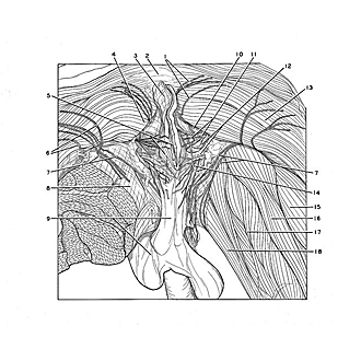
Dissection of anal triangle and ischiorectal fossae

General view of male perineum and ischiorectal fossae

Image #173-4

KEYWORDS: Muscles and tendons.

Creative Commons

Stanford holds the copyright to the David L. Bassett anatomical images and has assigned Creative Commons license Attribution-Share Alike 4.0 International to all of the images.

Creative Commons License logo

For additional information regarding use and permissions, please contact Dr. Drew Bourn at dbourn@stanford.edu.

Dissection of anal triangle and ischiorectal fossae
General view of male perineum and ischiorectal fossae
The membranous layer of superficial fossae (Colles' fascia) has been preserved in the urogenital triangle. In the anal triangle the ischiorectal fossae (10) have been dissected and the anal sphincter has been exposed. The irregular lunate fascia (12) has been preserved in the fossae and it is apparent that there is a continuity between this layer and Colles' fascia anteriorly. The left thigh has been sectioned slightly below the level of the trochanters of the femur.
1 . Medial cluneal nerves
2 . Coccyx (covered by periosteum and ligaments)
3 . Anococcygeal ligament
4 . Gluteus maximus muscle (portion arising from sacrotuberous ligament)
5 . Upper pointer: Anus Lower pointer: External anal sphincter muscle
6 . Inferior cluneal nerves
7 . Posterior perineal branches femoral cutaneous nerve
8 . Fascia lata
9 . Upper pointer: Superficial perineal fascia (Colles' fascia) Lower pointer: Scrotum
10 . Ischiorectal fossa (pointer indicates posterior recess of fossa)
11 . Left pointer: Inferior rectal nerves Right pointer: Inferior rectal artery
12 . Upper pointer: Lunate fascia Lower pointer: Posterior border of urogenital triangle
13 . Gluteus maximus muscle
14 . Posterior scrotal nerves
15 . Semimembranosus muscle
16 . Semitendinosus muscle
17 . adductor magnus muscle
18 . Gracilis muscle