Bassett Collection of Stereoscopic Images of Human Anatomy
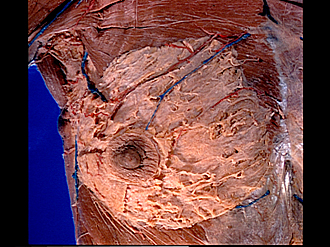
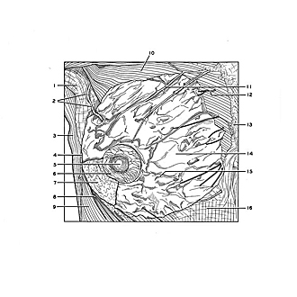
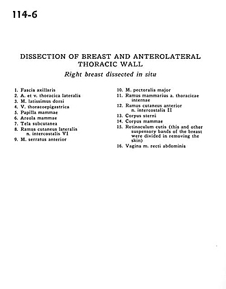
Dissection of breast and anterolateral thoracic wall
Right breast dissected in situ
Image #114-6
KEYWORDS: Breast.
Creative Commons
Stanford holds the copyright to the David L. Bassett anatomical images and has assigned Creative Commons license Attribution-Share Alike 4.0 International to all of the images.

For additional information regarding use and permissions, please contact the Medical History Center.
Dissection of breast and anterolateral thoracic wall
Right breast dissected in situ
- Axillary fascia
- Lateral thoracic artery and vein
- Latissimus dorsi muscle
- Thoracoepigastric vein
- Nipple (mammary papilla)
- Areola
- Superficial fascia
- Lateral cutaneous branch intercostal nerve VI
- Serratus anterior muscle
- Pectoralis major muscle
- Mammary branch internal thoracic artery
- Anterior cutaneous branch intercostal nerve II
- Body of sternum
- Mammary body
- Cutaneous ligaments (this and other suspensory bands of the breast were divided in removing the skin)
- Sheath of rectus abdominis muscle