
Bassett Collection of Stereoscopic Images of Human Anatomy
Elbow joint
Capsule and ligaments, anterior view of right elbow
Image #100-2
KEYWORDS: Elbow, Fascia ligaments and tendons, Muscles and tendons.
Creative Commons
Stanford holds the copyright to the David L. Bassett anatomical images and has assigned Creative Commons license Attribution-Share Alike 4.0 International to all of the images.

For additional information regarding use and permissions, please contact the Medical History Center.
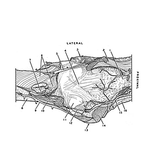

Elbow joint
Capsule and ligaments, anterior view of right elbow
- Body of humerus
- Brachialis muscle
- Lateral epicondyle of humerus
- Cubital joint capsule
- Recurrent radial artery
- Supinator muscle (deep part)
- Biceps brachii muscle (tendon of Insertion)
- Ulna
- Dorsal recurrent ulnar artery
- Brachialis muscle (insertion)
- Position of trochlea of humerus (covered by articular capsule)
- Branch anterior recurrent ulnar artery
- Medial epicondyle of humerus
- Pronator teres muscle (humeral head)
- Inferior Ulnar collateral artery
- Medial intermuscular septum