Shoulder
Capsule and ligaments of left shoulder joint, anterior view
Stanford holds the copyright to the David L. Bassett anatomical images and has assigned
Creative Commons license Attribution-Share
Alike 4.0 International to all of the images.
For additional information regarding use and permissions,
please contact the Medical History Center.
Image #94-3
Shoulder
Capsule and ligaments of left shoulder joint, anterior view
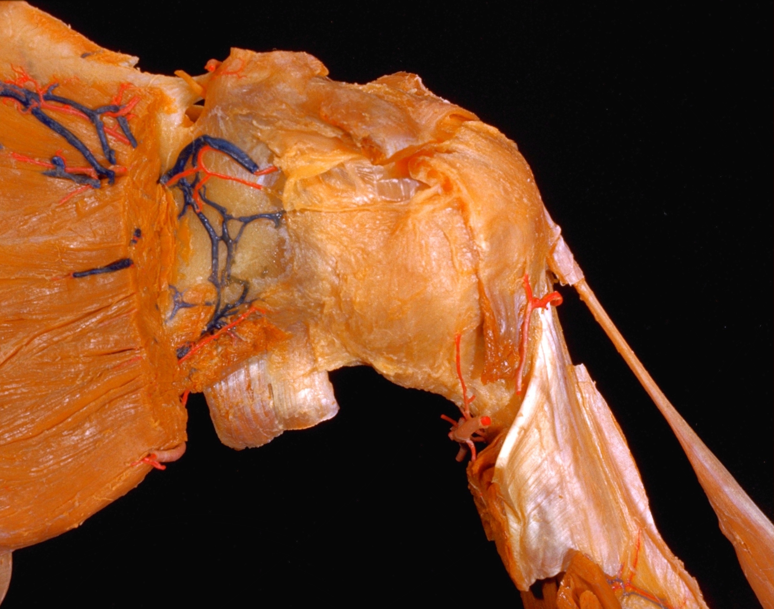
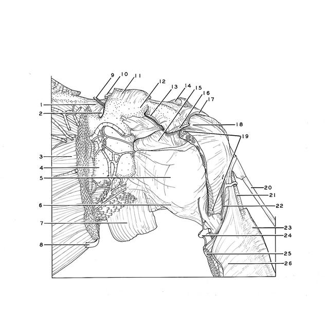
The subscapular muscle has been partially cut away from the specimen shown previously (91-7). The capsule of the shoulder joint is reinforced anteriorly by the middle and inferior glenohumeral ligaments (5,6). The contours of these extracapsular ligaments are not clearly visible externally. They are more evident in the interior of the joint (94-7).
- Superior transverse ligament of scapula
- Scapular notch
- Subscapularis muscle
- Subscapular fossa
- Joint capsule of humerus (pointer indicates position of middle glenohumeral ligament, visible internally in view 94-7)
- Joint capsule of humerus (pointer indicates position of inferior glenohumeral ligament, visible internally in view 94-7)
- Triceps brachii muscle (long head)
- Circumflex scapular artery
- Suprascapular nerve
- Transverse scapular artery
- Coracoclavicular ligament (cut off)
- Coracoid process of scapula
- Pectoralis minor muscle (tendon of insertion)
- Acromial articular surface
- Bursa of subscapularis muscle (translucent area of wall indicates position of a deeper bursal sac both bursae communicate with cavity of shoulder joint)
- Coracobrachialis muscle and short head bicipitis brachii muscle
- Coracoacromial ligament
- Coracohumeral ligament
- Tendon of subscapularis muscle (lower portion muscular)
- Biceps brachii muscle (long head, elevated)
- Body of humerus
- Anterior circumflex artery of humerus
- Pectoralis major muscle
- Posterior circumflex artery of humerus
- Teres major muscle
- Latissimus dorsi muscle