Dissection of head and neck from a posterior approach
Relations of internal jugular vein, hypoglossal, accessory and vagus nerves to atlantooccipital joint; atlantoepistrophic joint
Stanford holds the copyright to the David L. Bassett anatomical images and has assigned
Creative Commons license Attribution-Share
Alike 4.0 International to all of the images.
For additional information regarding use and permissions,
please contact the Medical History Center.
Image #80-5
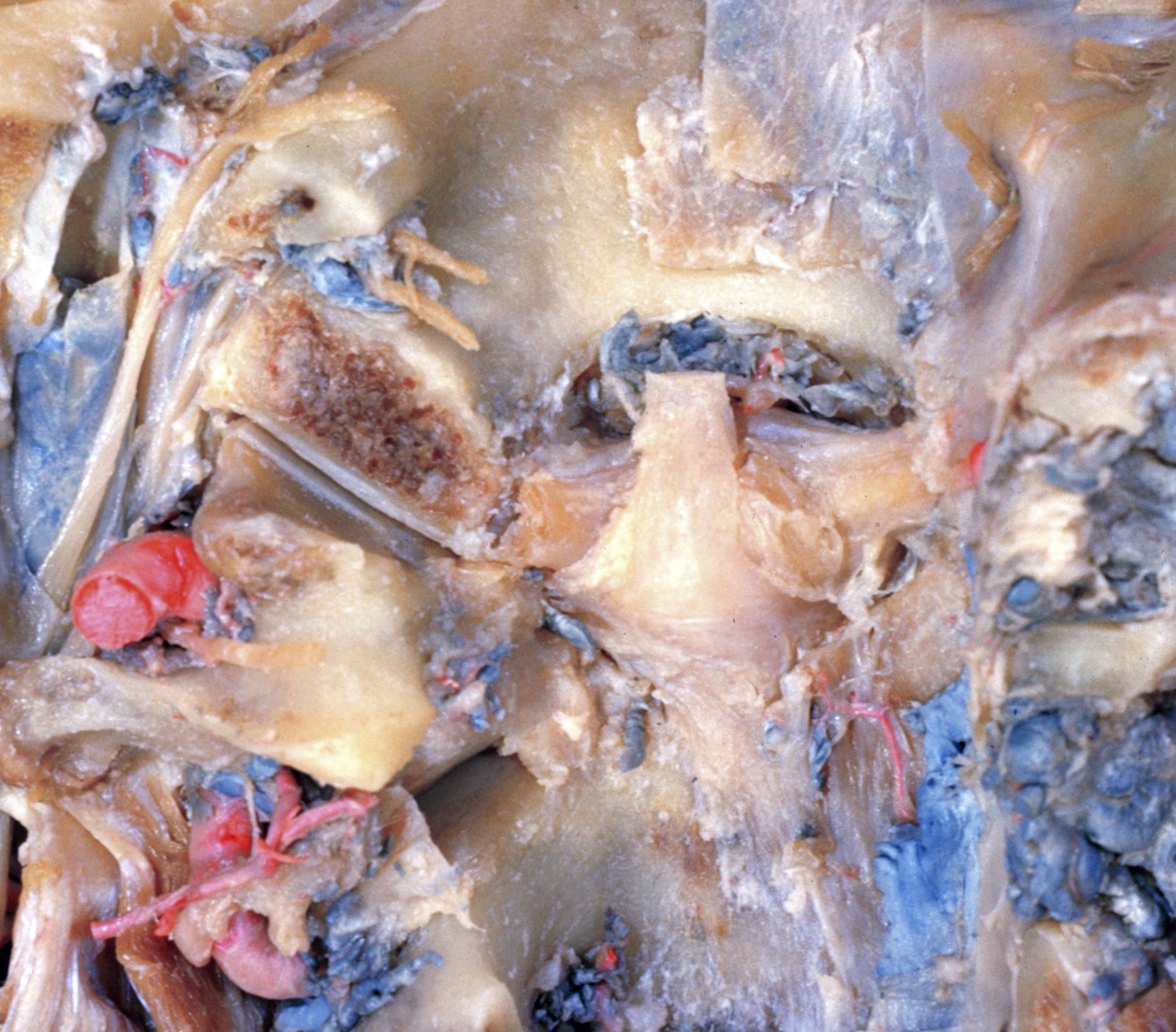
Dissection of head and neck from a posterior approach
Relations of internal jugular vein, hypoglossal, accessory and vagus nerves to atlantooccipital joint; atlantoepistrophic joint
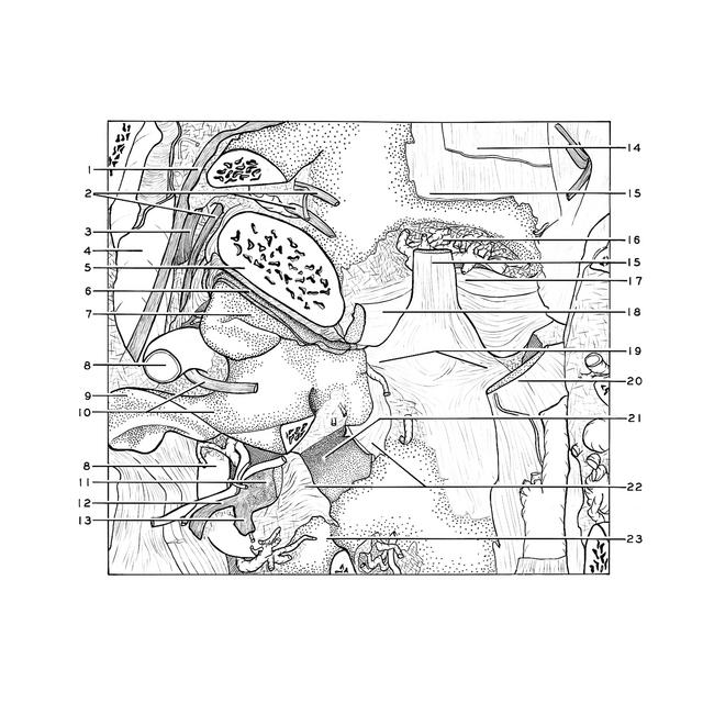
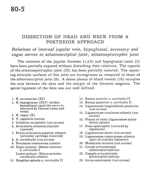
The contents of the jugular foramen (1,3,4) and hypoglossal canal (2) have been partially exposed without disturbing their relations. The capsule of the atlantoepistrophic joint (22) has been partially resected. The opposing articular surfaces of this joint are incongruous as compared to those of the atlantooccipital joint (6). A dense plexus of blood vessels (16) occupies the area between the dens and the margin of the foramen magnum. The apical ligament of the dens was not well defined.
- Accessory nerve (XI)
- Hypoglossal nerve (XII) (within hypoglossal canal the nerve is obscured by fibrous tissue and veins)
- Vagus nerve (X)
- Internal jugular vein
- Occipital condyle (cut across)
- Atlanto-occipital articulation (opened)
- Superior articular facet atlas (articular cartilage removed)
- Vertebral artery (cut across)
- Transverse process atlas
- Upper pointer: Anterior branch of cervical nerve I Lower pointer: Groove in atlas for vertebral artery
- Spinal ganglion cervical nerve II
- Anterior branch of cervical nerve II
- Posterior branch cervical nerve II
- Posterior longitudinal ligament (cut across)
- Cruciate ligament atlas (cut across)
- Plexus of veins (ligamentum apicis dentis absent)
- Dens axis (covered by ligaments)
- Alar ligament (cut across)
- Transverse ligament atlas (part of cruciate ligament)
- Tectorial membrane (cut across)
- Atlanto-occipital joint cavity
- Atlantoepistrophical joint capsule
- Arch of axis (cut across)