Exploration of the brain from its superior aspect
Corpus callosum and corona radiata
Stanford holds the copyright to the David L. Bassett anatomical images and has assigned
Creative Commons license Attribution-Share
Alike 4.0 International to all of the images.
For additional information regarding use and permissions,
please contact the Medical History Center.
Image #8-6
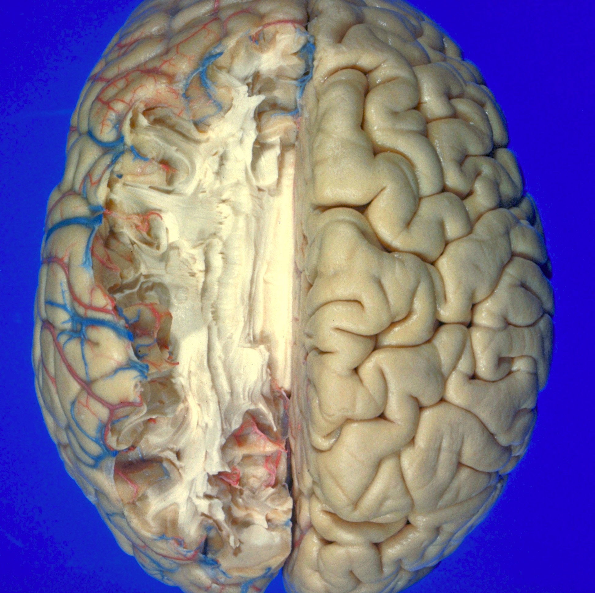
Exploration of the brain from its superior aspect
Corpus callosum and corona radiata
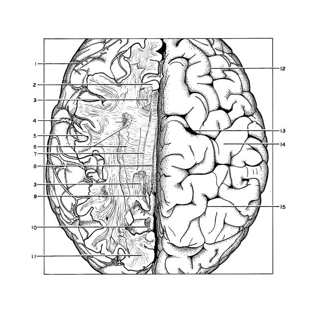
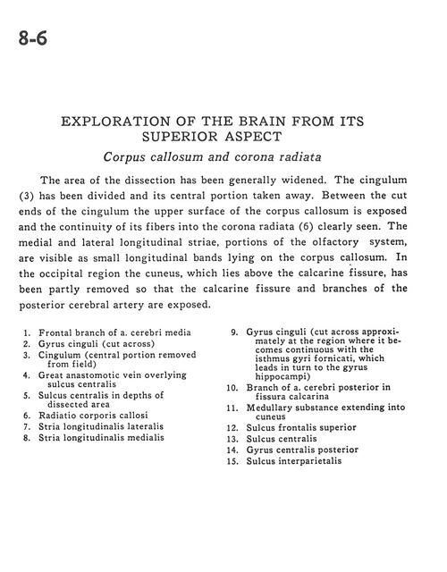
The area of the dissection has been generally widened. The cingulum (3) has been divided and its central portion taken away. Between the cut ends of the cingulum the upper surface of the corpus callosum is exposed and the continuity of its fibers into the corona radiata (6) clearly seen. The medial and lateral longitudinal striae, portions of the olfactory system, are visible as small longitudinal bands lying on the corpus callosum. In the occipital region the cuneus, which lies above the calcarine fissure, has been partly removed so that the calcarine fissure and branches of the posterior cerebral artery are exposed.
- Frontal branch of middle cerebral artery
- Cingulate gyrus (cut across)
- Cingulum (central portion removed from field)
- Great anastomotic vein overlying central sulcus
- Central sulcus in depths of dissected area
- Radiations corpus callosum
- Lateral longitudinal stria
- Medial longitudinal stria
- Cingulate gyrus (cut across approximately at the region where it becomes continuous with the Limbic lobe, which leads in turn to the hippocampal gyrus)
- Branch of posterior cerebral artery in calcarine fissure
- Medullary substance extending into cuneus
- Superior frontal sulcus
- Central sulcus
- Postcentral gyrus
- Interparietal sulcus