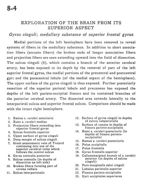
Exploration of the brain from its superior aspect
Gyrus cinguli; medullary substance of superior frontal gyrus
Stanford holds the copyright to the David L. Bassett anatomical images and has assigned
Creative Commons license Attribution-Share
Alike 4.0 International to all of the images.
For additional information regarding use and permissions,
please contact the Medical History Center.
Image #8-4
Exploration of the brain from its superior aspect
Gyrus cinguli; medullary substance of superior frontal gyrus
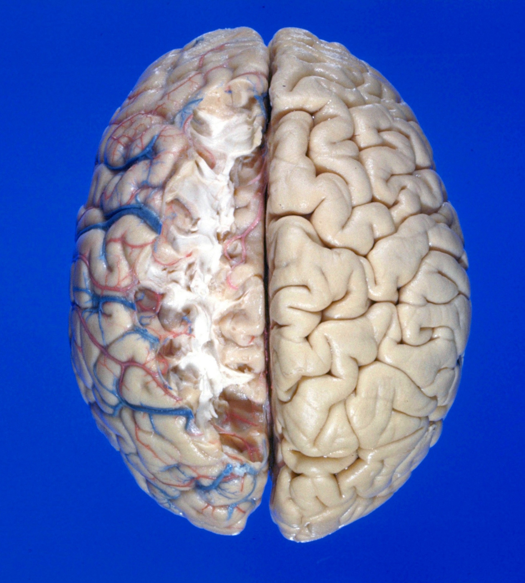
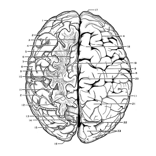
Medial portions of the left hemisphere have been removed to reveal systems of fibers in the medullary substance. In addition to short association fibers (arcuate fibers) the broken ends of longer association fibers and projection fibers are seen extending upward into the field of dissection. The sulcus cinguli (6), which contains a branch of the anterior cerebral artery, has been exposed to its depth by the removal of part of the left superior frontal gyrus, the medial portions of the precentral and postcentral gyri and the paracentral lobule (of the medial aspect of the hemisphere). The upper surface of the gyrus cinguli is thus exposed. Further posteriorly resection of the superior parietal lobule and precuneus has exposed the depths of the left parieto-occipital fissure and its contained branches of the posterior cerebral artery. The dissected area extends laterally to the interparietal sulcus and superior frontal sulcus. Comparison should be made with the intact right hemisphere.
- Branch anterior cerebral artery
- Branch middle cerebral artery
- Projection fibers extending into superior frontal gyrus
- Superior frontal sulcus
- Upper surface of cingulate gyrus
- Deep margin of cingulate sulcus
- Great anastomotic vein of Trolard continuing into one of the superior cerebral veins which follows the central sulcus
- Postcentral gyrus
- Central sulcus (in depths of dissection on left side)
- Broken fibers forming part of corona radiata
- Interparietal sulcus
- Surface of cingulate gyrus in depths of subparietal sulcus
- Surface of cuneus in depths of parieto-occipital fissure
- Branch posterior cerebral artery (in depths of parieto-occipital fissure)
- Branch posterior cerebral artery
- Occipital pole
- Frontal pole
- Superior frontal gyrus
- Callosomarginal branch of anterior cerebral artery (in depths of cingulate sulcus)
- Marginal part of cingulate sulcus
- Superior parietal lobule
- Parieto-occipital fissure
- Superior occipital gyri