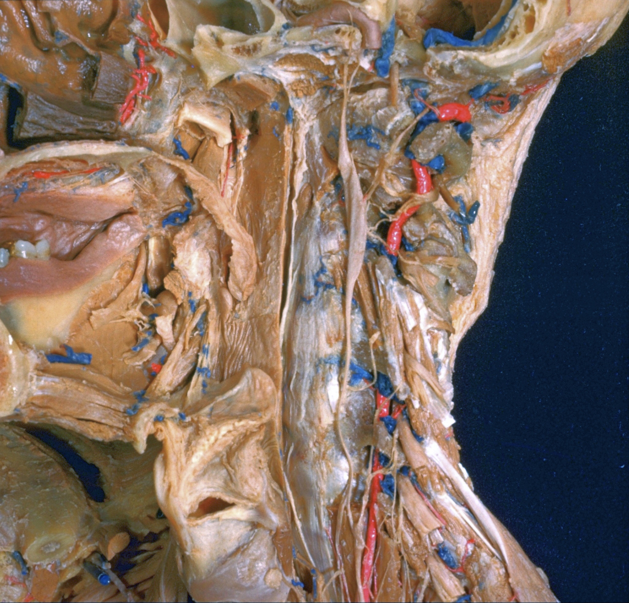
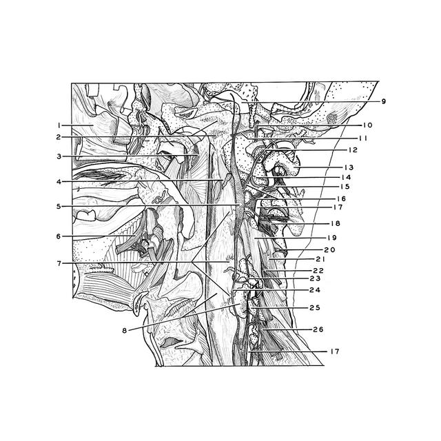
Dissection of anterior and lateral regions of neck
Vertebral artery; gray communicating rami of cervical sympathetic trunk, left anterolateral view
Stanford holds the copyright to the David L. Bassett anatomical images and has assigned
Creative Commons license Attribution-Share
Alike 4.0 International to all of the images.
For additional information regarding use and permissions,
please contact the Medical History Center.
Image #78-4
Dissection of anterior and lateral regions of neck
Vertebral artery; gray communicating rami of cervical sympathetic trunk, left anterolateral view
The longus colli, rectus capitis anterior, rectus capitis lateralis and some of the intertransverse muscles have been cut away. In addition, the transverse processes of the atlas, epistropheus and fifth cervical vertebra have been cut so that the course of the vertebral artery (17) is visible. The artery did not pass through the transverse process of the sixth cervical vertebra in this specimen.
- Nasal cavity (for details refer to reel 72-3)
- Area of insertion of longus capitis muscle
- Nasal part pharynx (for details refer to reel 78-5)
- Longus colli muscle (transected close to insertion on anterior tubercle atlas)
- Superior cervical ganglion
- Lingual nerve
- Areas of insertion of Iongus colli muscle on bodies of cervical vertebraes III-V
- Upper pointer: Position of intervertebral disc uniting cervical vertebrae IV-V Lower pointer: Body of cervical vertebra V
- Internal carotid artery
- Hypoglossal nerve (XII) near hypoglossal canal
- Atlanto-occipital articulation
- Anterior branch of cervical nerve I (branch communicating with hypoglossal nerve cut away)
- Posterior arch of atlas
- Articulation of atlas-axis
- Anterior branch of cervical nerve II
- Gray rami communicantes
- Vertebral artery
- Cervical nerve III
- Intertransverse muscle (anterior)
- Posterior branch cervical nerve IV
- Intertransverse muscle (posterior)
- Anterior branch of cervical nerve IV
- Gray rami communicans
- Sympathetic trunk
- Anterior tubercle cervical vertebra V
- Cervical nerve VI