Dissection of anterior and lateral regions of neck
Larynx; relation to oral cavity, oral and laryngeal parts of pharynx, left lateral view
Stanford holds the copyright to the David L. Bassett anatomical images and has assigned
Creative Commons license Attribution-Share
Alike 4.0 International to all of the images.
For additional information regarding use and permissions,
please contact the Medical History Center.
Image #78-1
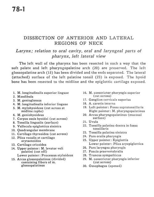
Dissection of anterior and lateral regions of neck
Larynx; relation to oral cavity, oral and laryngeal parts of pharynx, left lateral view
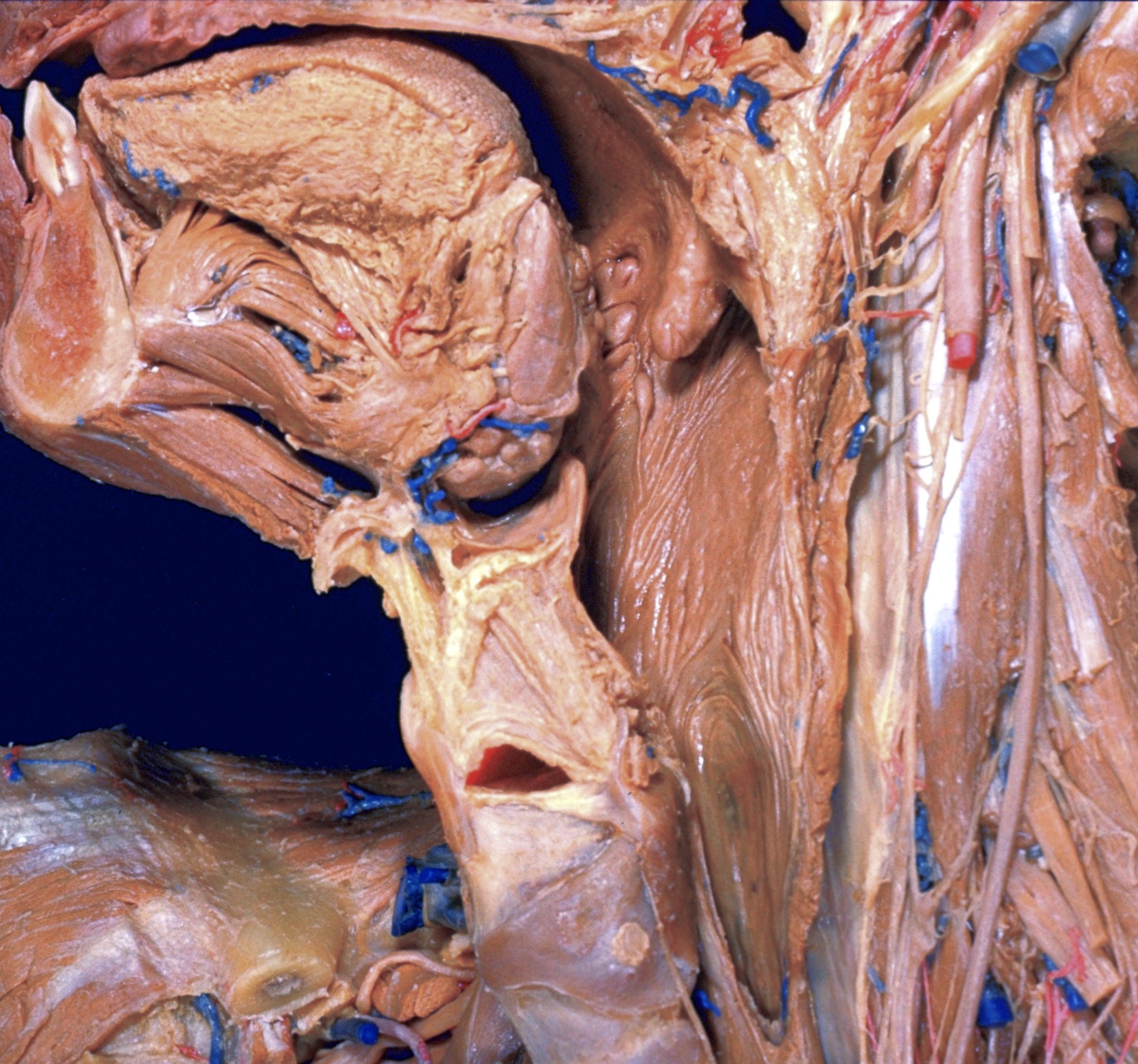
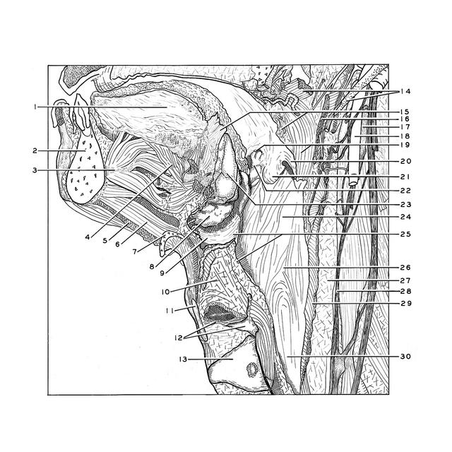
The left wall of the pharynx has been resected in such a way that the soft palate and left pharyngopalatine arch (20) are preserved. The left glossopalatine arch (15) has been divided and the ends separated. The lateral (attached) surface of the left palatine tonsil (23) is exposed. The hyoid bone has been resected to the midline and the epiglottic cartilage exposed.
- Superior longitudinal muscle of tongue
- Mandible
- Genioglossus muscle
- Inferior longitudinal muscle of tongue
- Mylohyoid muscle (cut across at midline raphe)
- Geniohyoid muscle
- Body hyoid bone (cut across)
- Lingual tonsil (surface)
- Vallecula epiglottica left
- Quadrangular membrane
- Thyroid cartilage (cut across)
- Vocal cord and arytenoid cartilage
- Cricoid cartilage
- Upper pointer: Levator veli palatini muscle (cut off) Lower pointer: Styloid process
- Palatoglossal arch (divided) containing fibers of glossopalatine muscle
- Superior pharyngeal constrictor muscle (cut across)
- Superior cervical ganglion
- Internal carotid artery
- Left pointer: Supratonsillar fossa Right pointer: Pharyngopalatine muscle
- Palatopharyngeal arch (mucosal surface)
- Uvula
- Palatine tonsil right in tonsillar fossa
- Palatine tonsil left
- Oral part pharynx
- Upper pointer: Epiglottis Lower pointer: Aryepiglottic fold
- Laryngeal part of pharynx
- Prevertebral fascia
- Sympathetic trunk
- Inferior pharyngeal constrictor muscle (cut across)
- Esophagus (opened)