Dissection of anterior and lateral regions of neck
Left lobe of thyroid gland in situ, anterior view
Stanford holds the copyright to the David L. Bassett anatomical images and has assigned
Creative Commons license Attribution-Share
Alike 4.0 International to all of the images.
For additional information regarding use and permissions,
please contact the Medical History Center.
Image #74-6
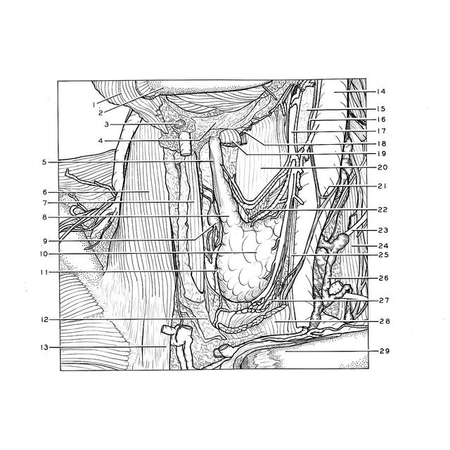
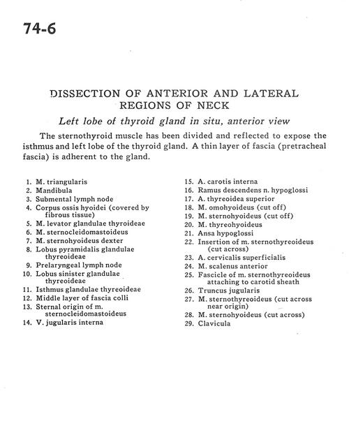
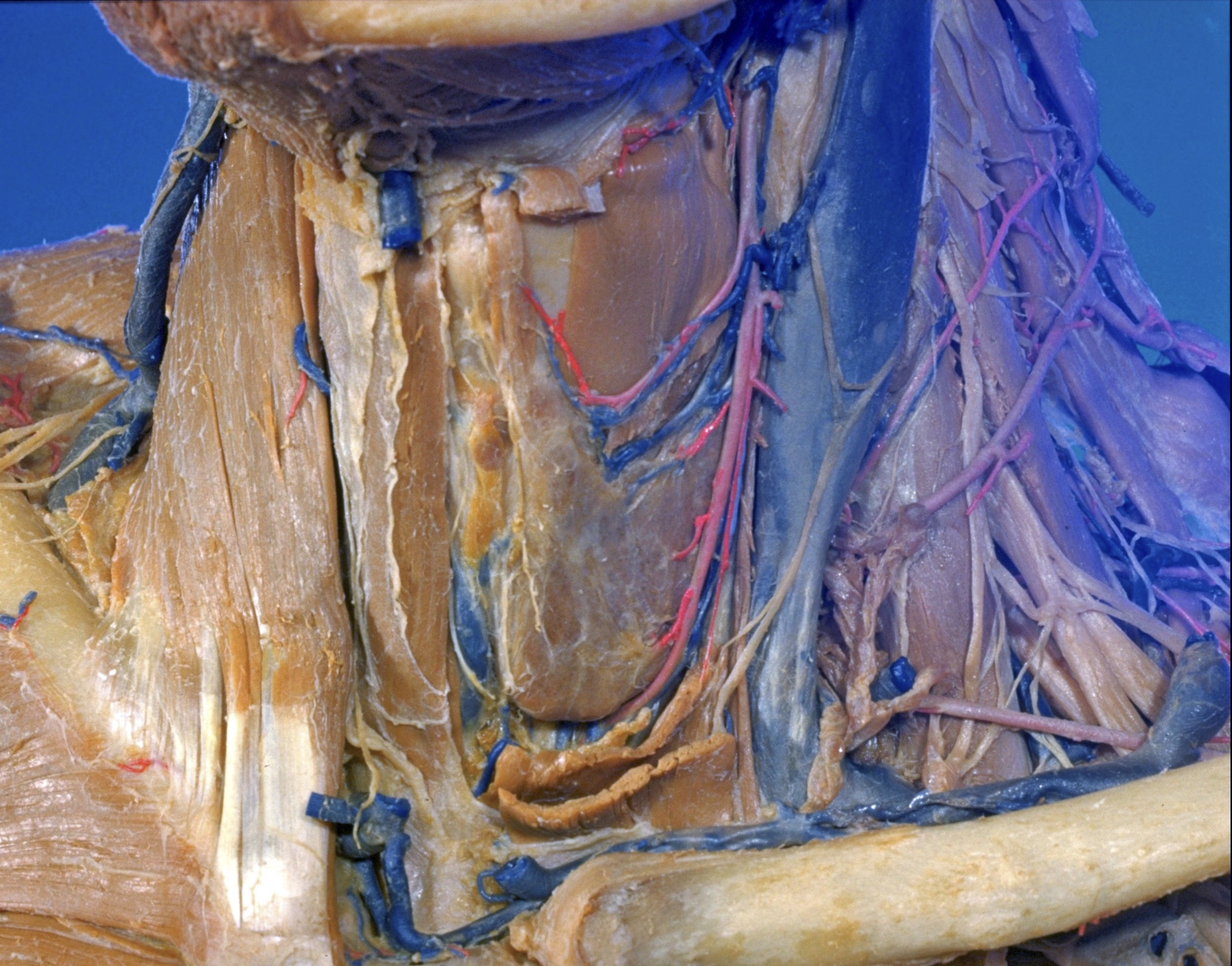
Dissection of anterior and lateral regions of neck
Left lobe of thyroid gland in situ, anterior view
The sternothyroid muscle has been divided and reflected to expose the isthmus and left lobe of the thyroid gland. A thin layer of fascia (pretracheal fascia) is adherent to the gland.
- Depressor anguli oris muscle
- Mandible
- Submental lymph node
- Body hyoid bone (covered by fibrous tissue)
- Levator muscle of thyroid gland
- Sternocleidomastoid muscle
- Sternohyoid muscle right
- Pyramidal lobe of thyroid gland
- Prelaryngeal lymph node
- Left lobe of thyroid gland
- Isthmus of thyroid gland
- Middle layer of superficial fascia
- Sternal origin of sternocleidomastoid muscle
- Internal jugular vein
- Internal carotid artery
- Descending branch hypoglossal nerve
- Superior thyroid artery
- Omohyoid muscle (cut off)
- Sternohyoid muscle (cut off)
- Thyrohyoid muscle
- Ansa hypoglossi
- Insertion of sternothyroid muscle (cut across)
- Superficial cervical artery
- Anterior scalene muscle
- Fascicle of sternothyroid muscle attaching to carotid sheath
- Jugular trunk
- Sternothyroid muscle (cut across near origin)
- Sternohyoid muscle (cut across)
- Clavicle