Dissection of anterior and lateral regions of neck
Relation of external and middle layers of cervical fascia to carotid sheath, anterolateral view
Stanford holds the copyright to the David L. Bassett anatomical images and has assigned
Creative Commons license Attribution-Share
Alike 4.0 International to all of the images.
For additional information regarding use and permissions,
please contact the Medical History Center.
Image #73-3
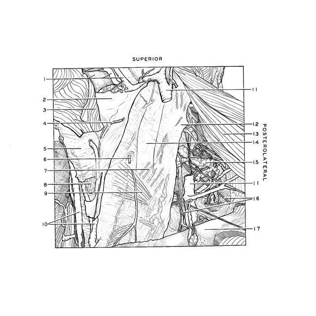
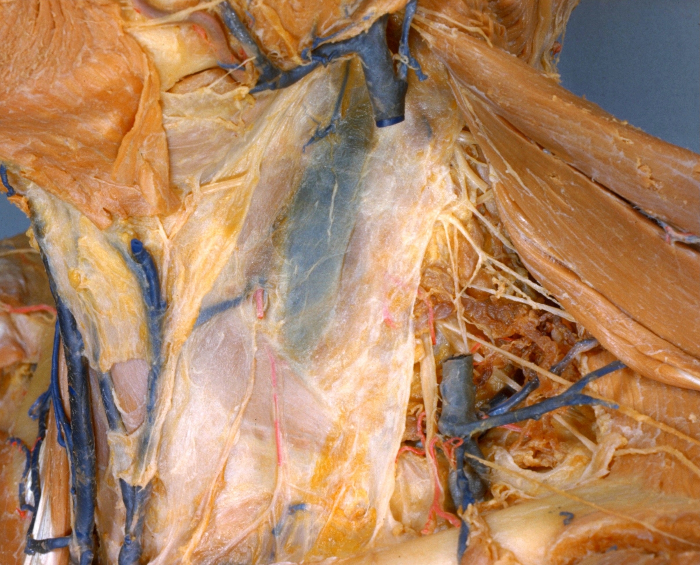
Dissection of anterior and lateral regions of neck
Relation of external and middle layers of cervical fascia to carotid sheath, anterolateral view
The sternocleidomastoid muscle (13) has been cut from its origins and reflected laterally. The deep lamina of the external layer of cervical fascia, which covered the deep surface of the muscle, has been retained. This fascia is intimately related to the middle layer of cervical fascia which covers the omohyoid muscle (9), as well as to the part of the carotid sheath which encloses the internal jugular vein (14).
- Anterior facial vein and external maxillary artery (passing superiorly across mandible)
- Cervical fascia investing submaxillary gland
- Platysma (reflected anteriorly)
- Cutaneus colli nerve (cut off)
- Tela subcutanea in midline
- Sternocleidomastoid branch of superior thyroid artery
- Profunda lymphatic vessel (amber colored streaks within fascia)
- Sternohyoid muscle (covered by middle layer of cervical fascia)
- Omohyoid muscle (covered by deep lamina of external layer of cervical fascia in addition to middle layer of cervical fascia)
- Anterior jugular veins
- External jugular vein (cut off)
- Cervical fascia (deep lamina of external layer)
- Sternocleidomastoid muscle (reflected laterally)
- Internal jugular vein (covered by carotid sheath)
- Superficial cervical lymph gland
- Supraclavicular nerves
- Clavicle
- [Legend above restored translation from Latin]