Dissection of anterior and lateral regions of neck
Suprasternal space, anterior view
Stanford holds the copyright to the David L. Bassett anatomical images and has assigned
Creative Commons license Attribution-Share
Alike 4.0 International to all of the images.
For additional information regarding use and permissions,
please contact the Medical History Center.
Image #72-6
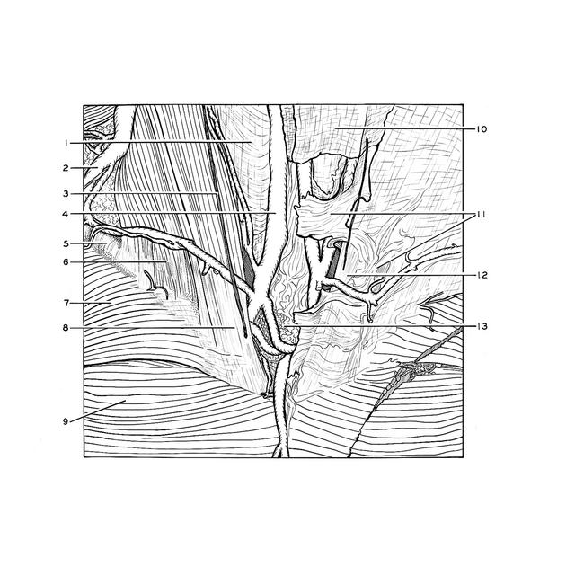
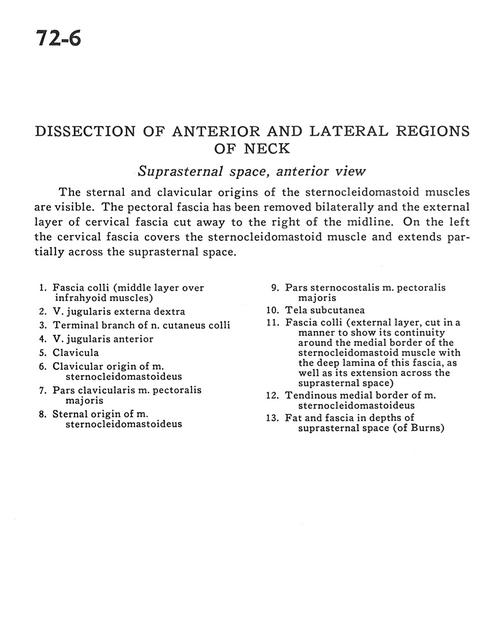
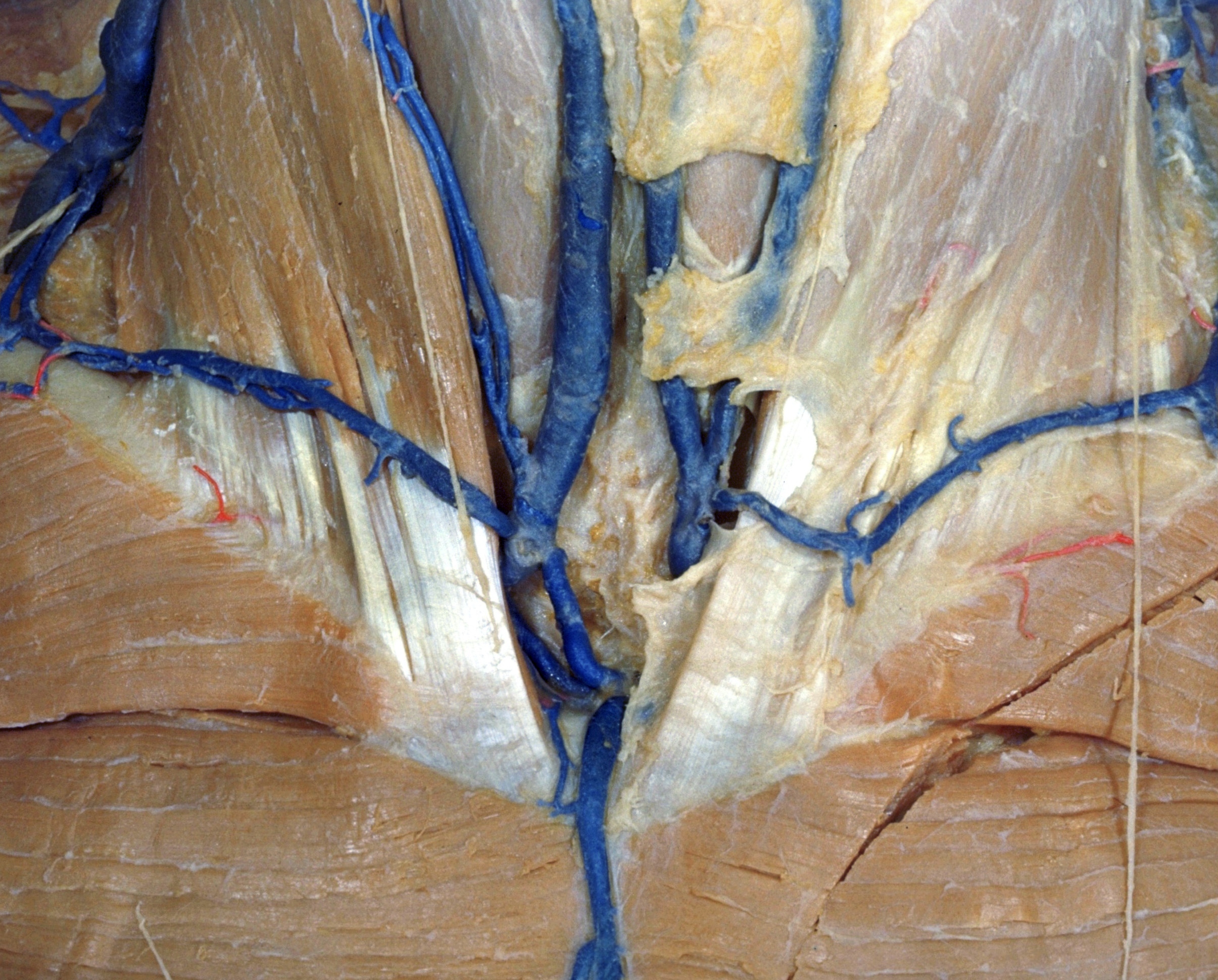
Dissection of anterior and lateral regions of neck
Suprasternal space, anterior view
The sternal and clavicular origins of the sternocleidomastoid muscles are visible. The pectoral fascia has been removed bilaterally and the external layer of cervical fascia cut away to the right of the midline. On the left the cervical fascia covers the sternocleidomastoid muscle and extends partially across the suprasternal space.
- Fascia colli (middle layer over infrahyoid muscles)
- Right external jugular vein
- Terminal branch of cutaneus colli nerve
- Anterior jugular vein
- Clavicula
- Clavicular origin of sternocleidomastoid muscle
- Clavicular part of pectoralis major muscle
- Sternal origin of sternocleidomastoid muscle
- Sternocostal part of pectoralis major muscle
- Tela subcutanea
- Fascia colli (external layer, cut in a manner to show its continuity around the medial border of ther sternocleidomastoid muscle with the deep lamina of this fascia, as well as its extension
- Tendinous medial border of sternocleidomastoid muscle
- Fat and fascia in depths of suprasternal space (of Burns)
- [Legend above restored translation from Latin]