Dissection of nasal fossae, nasal pharynx, and palate
Relation of nasal septum to sphenoid sinus and hypophysis; right palatine nerves; palatine arches, left lateral view
Stanford holds the copyright to the David L. Bassett anatomical images and has assigned
Creative Commons license Attribution-Share
Alike 4.0 International to all of the images.
For additional information regarding use and permissions,
please contact the Medical History Center.
Image #72-1
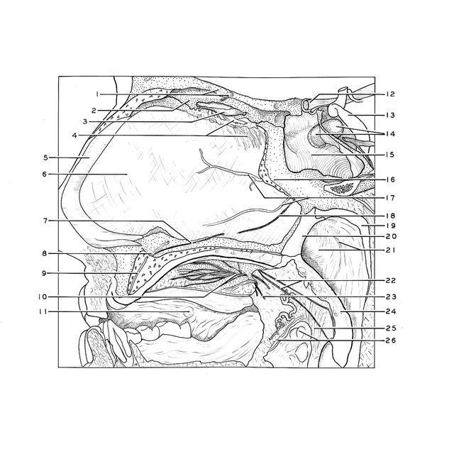
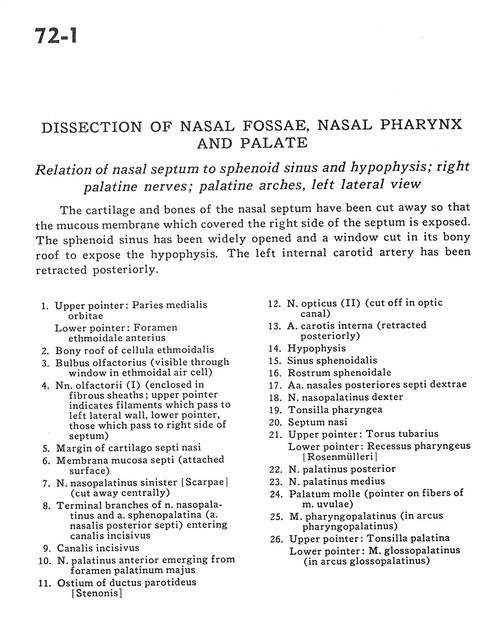
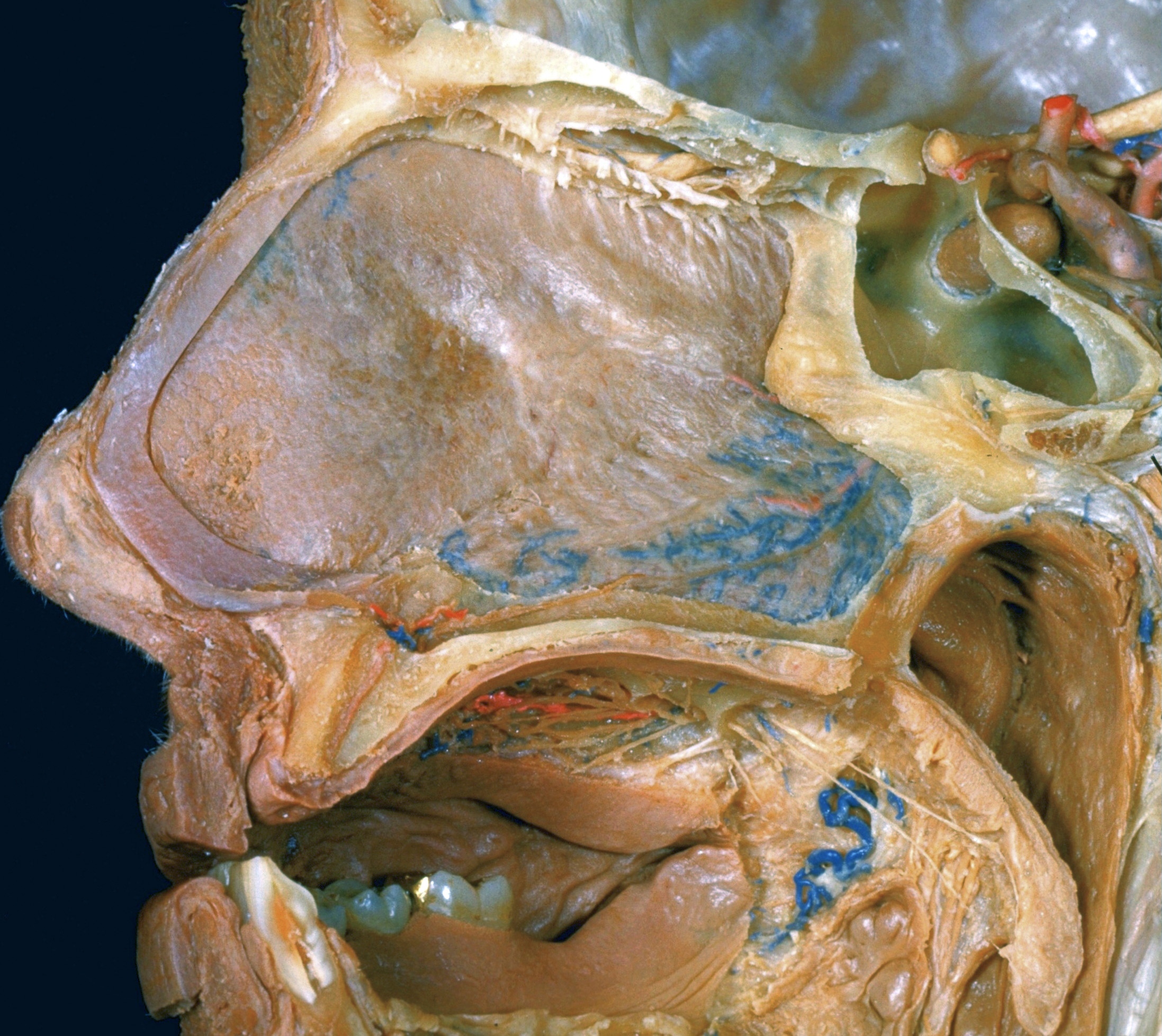
Dissection of nasal fossae, nasal pharynx, and palate
Relation of nasal septum to sphenoid sinus and hypophysis; right palatine nerves; palatine arches, left lateral view
The cartilage and bones of the nasal septum have been cut away so that the mucous membrane which covered the right side of the septum is exposed. The sphenoid sinus has been widely opened and a window cut in its bony roof to expose the hypophysis. The left internal carotid artery has been retracted posteriorly.
- Upper pointer: Medial wall of orbit Lower pointer: Anterior ethmoidal foramen
- Bony roof of ethmoidal cell
- Olfactory bulb (visible through window in ethmoidal air cell)
- Olfactory nerve (I) (enclosed in fibrous sheaths; upper pointer indicates filaments which pass to left lateral wall, lower pointer those which pass to right side of septum)
- Margin of septal cartilage
- Mucosal membrane septi (attached surface)
- Nasopalatine nerve left (cut away centrally)
- Terminal branches of nasopalatine nerve and sphenopalatine artery (posterior septal artery) entering incisive canal
- Incisive canal
- Anterior palatine nerve emerging from greater palatine foramen
- Ostium of parotid duct
- Optic nerve (II) (cut off in optic canal)
- Internal carotid artery (retracted posteriorly)
- Hypophysis
- Sphenoid sinus
- Rostrum of sphenoid
- Posterior nasal septal arteries right
- Nasopalatine nerve right
- Pharyngeal tonsil
- Nasal septum
- Upper pointer: Torus tubarius Lower pointer: Pharyngeal recess
- Posterior palatine nerve
- Middle palatine nerve
- Soft palate (pointer on fibers of uvular muscle)
- Pharyngopalatine muscle (in palatopharyngeal arch)
- Upper pointer: Palatine tonsil Lower pointer: Glossopalatine muscle (in palatoglossal arch)