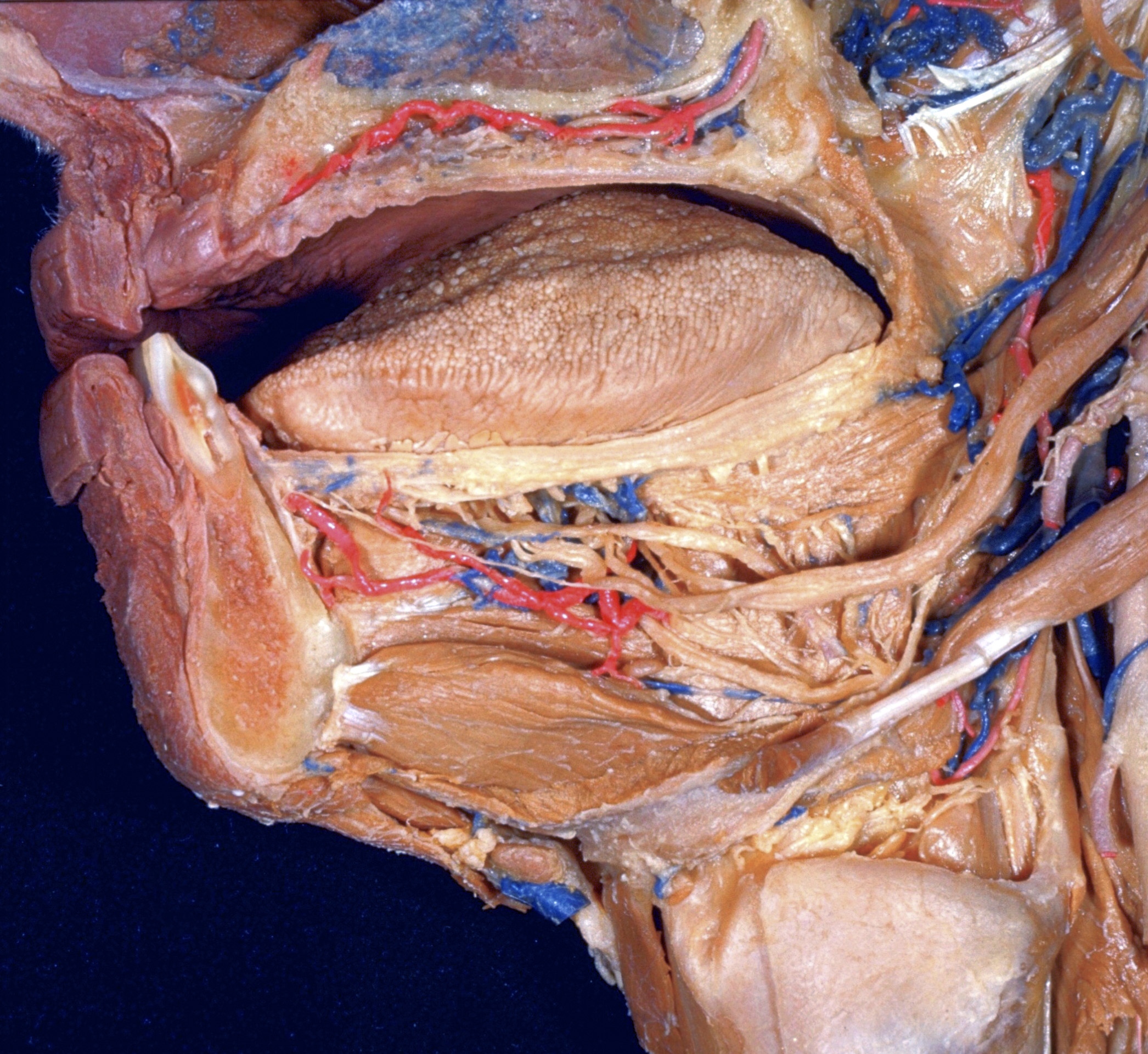
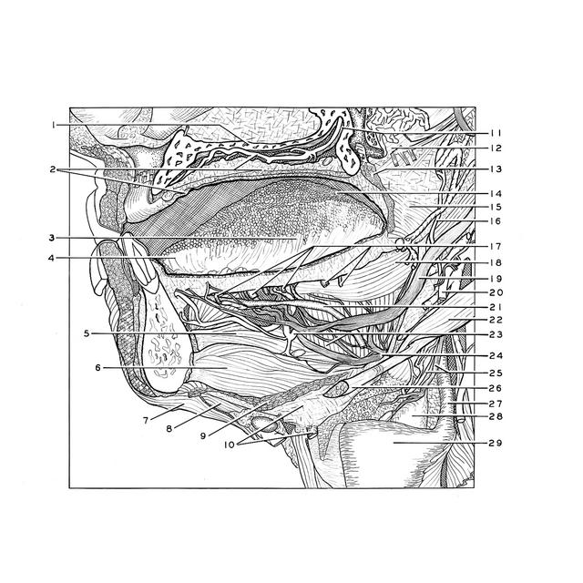
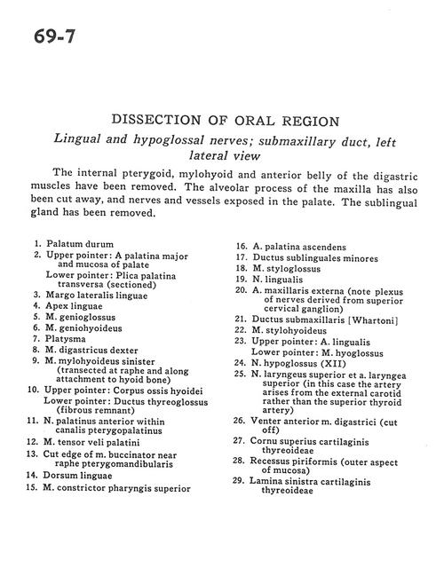
Dissection of oral region
Lingual and hypoglossal nerves; submaxillary duct, left lateral view
Stanford holds the copyright to the David L. Bassett anatomical images and has assigned
Creative Commons license Attribution-Share
Alike 4.0 International to all of the images.
For additional information regarding use and permissions,
please contact the Medical History Center.
Image #69-7
Dissection of oral region
Lingual and hypoglossal nerves; submaxillary duct, left lateral view
The internal pterygoid, mylohyoid and anterior belly of the digastric muscles have been removed. The alveolar process of the maxilla has also been cut away, and nerves and vessels exposed in the palate. The sublingual gland has been removed.
- Hard palate
- Upper pointer: Greater palatine artery and mucosa of palate Lower pointer: Transverse palatine fold (sectioned)
- Lateral lingual margin
- Apex of tongue
- Genioglossus muscle
- Geniohyoid muscle
- Platysma
- Digastric muscle right
- Mylohyoid muscle left (transected at raphe and along attachment to hyoid bone)
- Upper pointer: Body of hyoid bone Lower pointer: Thyroglossal duct (fibrous remnant)
- Anterior palatine nerve within pterygopalatine canal
- Tensor veli palatini muscle
- Cut edge of buccinator muscle near pterygomandibular raphe
- Dorsum of tongue
- Superior pharyngeal constrictor muscle
- Ascending palatine artery
- Minor sublingual duct
- Styloglossus muscle
- Lingual nerve
- External maxillary artery (note plexus of nerves derived from superior cervical ganglion)
- Submandibular duct
- Stylohyoid muscle
- Upper pointer: Lingual artery Lower pointer: Hyoglossus muscle
- Hypoglossal nerve (XII)
- Superior laryngeal nerve and superior laryngeal artery (in this case the artery arises from the external carotid rather than the superior thyroid artery)
- Anterior belly digastric muscle (cut off)
- Superior horn thyroid cartilage
- Piriform recess (outer aspect of mucosa)
- Left lamina thyroid cartilage