Dissection of oral region
Nerves and blood vessels to upper teeth, left lateral view
Stanford holds the copyright to the David L. Bassett anatomical images and has assigned
Creative Commons license Attribution-Share
Alike 4.0 International to all of the images.
For additional information regarding use and permissions,
please contact the Medical History Center.
Image #69-2
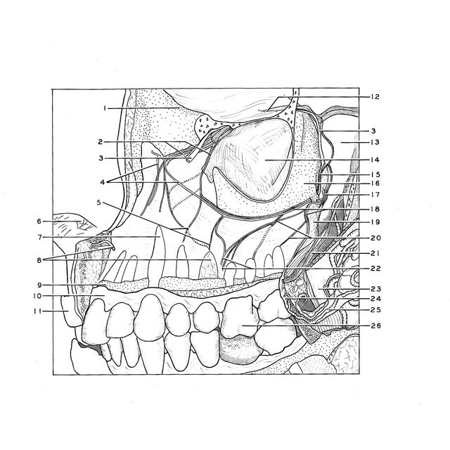
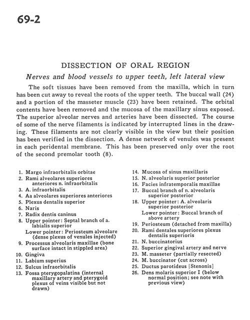
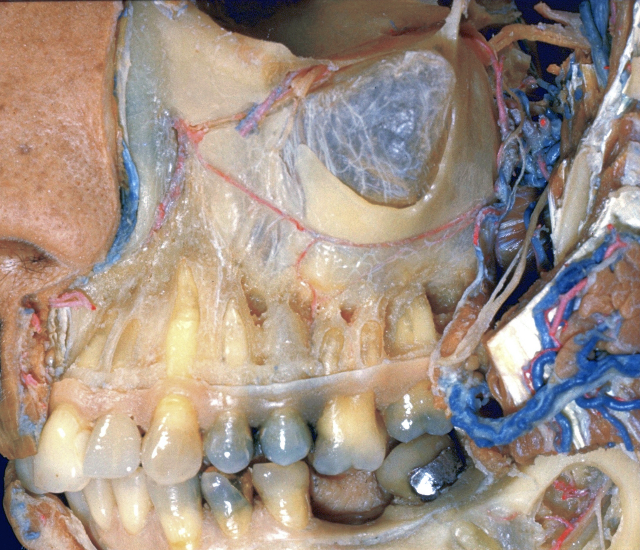
Dissection of oral region
Nerves and blood vessels to upper teeth, left lateral view
The soft tissues have been removed from the maxilla, which in turn has been cut away to reveal the roots of the upper teeth. The buccal wall (24) and a portion of the masseter muscle (23) have been retained. The orbital contents have been removed and the mucosa of the maxillary sinus exposed. The superior alveolar nerves and arteries have been dissected. The course of some of the nerve filaments is indicated by interrupted lines in the drawing. These filaments are not clearly visible in the view but their position has been verified in the dissection. A dense network of venules was present in each peridental membrane. This has been preserved only over the root of the second premolar tooth (8).
- Infraorbital margin
- Anterior superior alveolar branches infraorbital nerve
- Infraorbital artery
- Anterior superior alveolar arteries
- Superior dental plexus
- Naris
- Root of canine
- Upper pointer: Septal branch of superior labial artery Lower pointer: Alveolar periosteum (dense plexus of venules injected)
- Alveolar process of maxilla (bone surface intact in stippled area)
- Gingiva
- Upper lip
- Infraorbital sulcus
- Pterygopalatine fossa (internal maxillary artery and pterygoid plexus of veins visible but not drawn)
- Mucosa of maxillary sinus
- Superior posterior alveolar nerve
- Infratemporal surface of maxilla
- Buccal branch of superior posterior alveolar nerve
- Upper pointer: Superior posterior alveolar artery Lower pointer: Buccal branch of above artery
- Periosteum (detached from maxilla)
- Superior dental branches of superior dental plexus
- Buccal nerve
- Superior gingival artery and nerve
- Masseter muscle (partially resected)
- Buccinator muscle (cut across)
- Parotid duct
- Upper molar I (below normal position see note with previous view)