Dissection of left infratemporal and pterygopalatine fossae
Nerve supply to internal pterygoid muscle; mandibular division of trigeminal nerve, lateral view
Stanford holds the copyright to the David L. Bassett anatomical images and has assigned
Creative Commons license Attribution-Share
Alike 4.0 International to all of the images.
For additional information regarding use and permissions,
please contact the Medical History Center.
Image #66-1
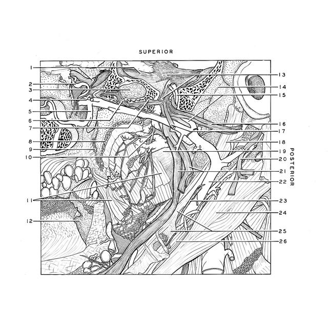
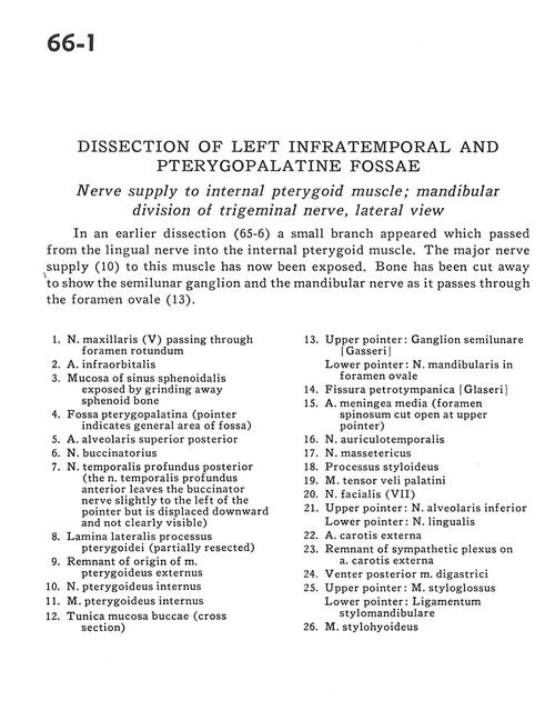
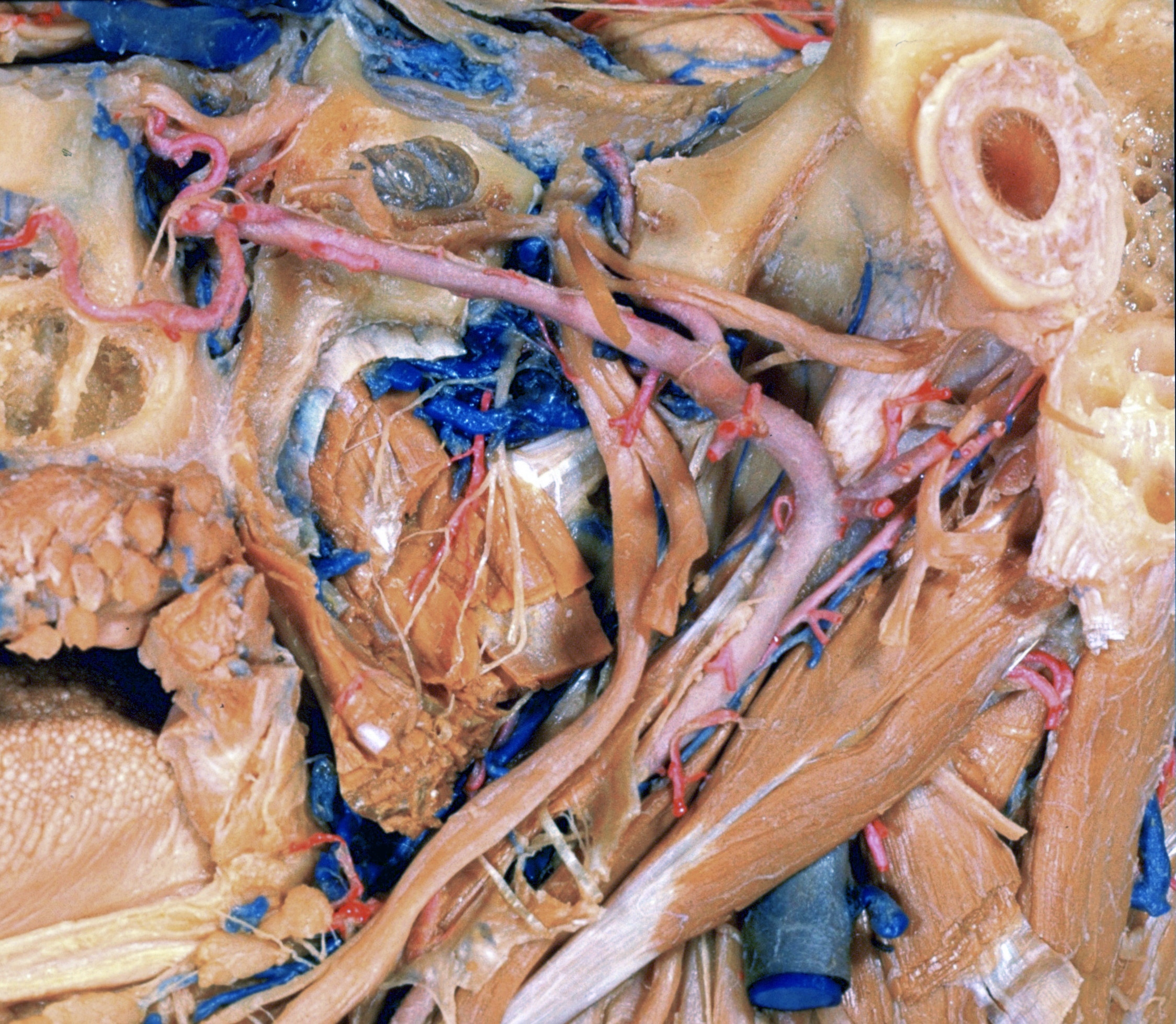
Dissection of left infratemporal and pterygopalatine fossae
Nerve supply to internal pterygoid muscle; mandibular division of trigeminal nerve, lateral view
In an earlier dissection (65-6) a small branch appeared which passed from the lingual nerve into the internal pterygoid muscle. The major nerve supply (10) to this muscle has now been exposed. Bone has been cut away to show the semilunar ganglion and the mandibular nerve as it passes through the foramen ovale (13).
- Maxillary nerve (V) passing through foramen rotundum
- Infraorbital artery
- Mucosa of sphenoid sinus exposed by grinding away sphenoid bone
- Pterygopalatine fossa (pointer indicates general area of fossa)
- Superior posterior alveolar artery
- Buccinator nerve
- Posterior deep temporal nerve (the anterior deep temporal nerve leaves the buccinator nerve slightly to the left of the pointer but is displaced downward and not clearly visible)
- Lateral plate of pterygoid process (partially resected)
- Remnant of origin of external pterygoid muscle
- Internal pterygoid nerve
- Internal pterygoid muscle
- Buccal mucosa (cross section)
- Upper pointer: Semilunar ganglion (trigeminal) Lower pointer: Mandibular nerve in foramen ovale
- Petrotympanic fissure
- Middle meningeal artery (foramen spinosum cut open at upper pointer)
- Auriculotemporal nerve
- Masseteric nerve
- Styloid process
- Tensor veli palatini muscle
- Facial nerve (VII)
- Upper pointer: Inferior alveolar nerve Lower pointer: Lingual nerve
- External carotid artery
- Remnant of sympathetic plexus on external carotid artery
- Posterior belly of digastric muscle
- Upper pointer: Styloglossus muscle Lower pointer: Stylomandibular ligament
- Stylohyoid muscle