Dissection of left infratemporal and pterygopalatine fossae
Cavity of temporomandibular articulation, inferolateral view
Stanford holds the copyright to the David L. Bassett anatomical images and has assigned
Creative Commons license Attribution-Share
Alike 4.0 International to all of the images.
For additional information regarding use and permissions,
please contact the Medical History Center.
Image #65-7
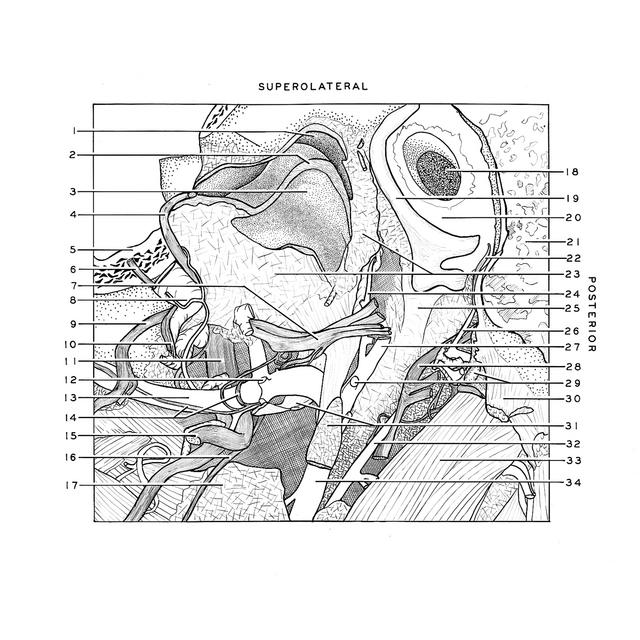
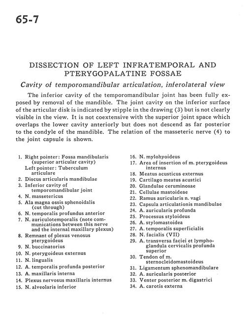
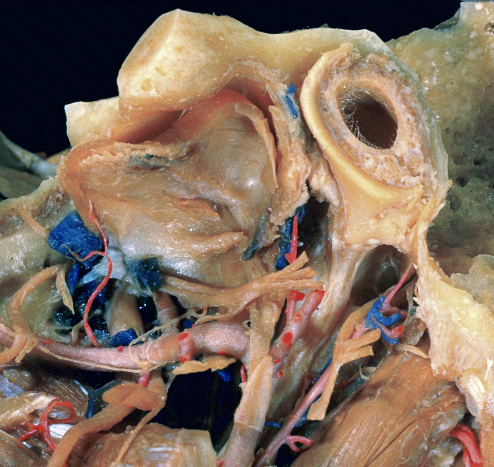
Dissection of left infratemporal and pterygopalatine fossae
Cavity of temporomandibular articulation, inferolateral view
The inferior cavity of the temporomandibular joint has been fully exposed by removal of mandible. The joint cavity on the inferior surface of the articular disk is indicated by stipple in the drawing (3) but is not clearly visible in the view. It is not coextensive with the superior joint space which overlaps the lower cavity anteriorly but does not descend as far as posterior to the condyle of the mandible. The relation of the masseteric nerve (4) to the joint capsule is shown.
- Right pointer: Mandibular fossa (superior articular cavity) Left pointer: Tubercle of articulation
- Articular disc of mandible
- Inferior cavity of temporomandibular joint
- Masseteric nerve
- Greater wing of sphenoid (cut through)
- Anterior deep temporal nerve
- Auriculotemporal nerve (note communications between this nerve and the internal maxillary plexus)
- Remnant of pterygoid venous plexus
- Buccal nerve
- External pterygoid nerve
- Lingual nerve
- Deep posterior temporal artery
- Internal maxillary artery
- Internal maxillary nerve plexus
- Inferior alveolar nerve
- Mylohyoid nerve
- Area of insertion of internal pterygoid muscle
- External acoustic meatus
- Cartilaginous acoustic meatus
- Ceruminous gland
- Mastoid cells
- Auricular branch vagus nerve
- Joint capsule of mandible
- Deep auricular artery
- Styloid process
- Stylomastoid artery
- Superficial temporal artery
- Facial nerve (VII)
- Transverse facial artery and superior deep cervical lymph node
- Tendon of sternocleidomastoid muscle
- Sphenomandibular ligament
- Posterior auricular artery
- Posterior belly of digastric muscle
- External carotid artery