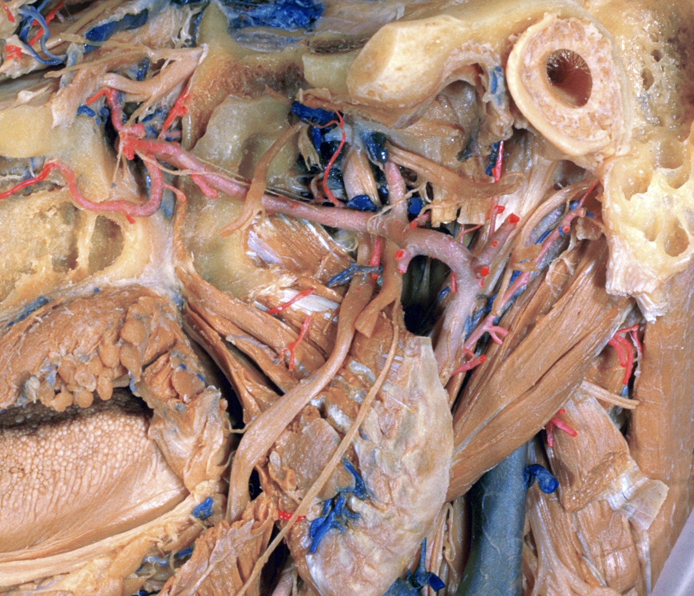
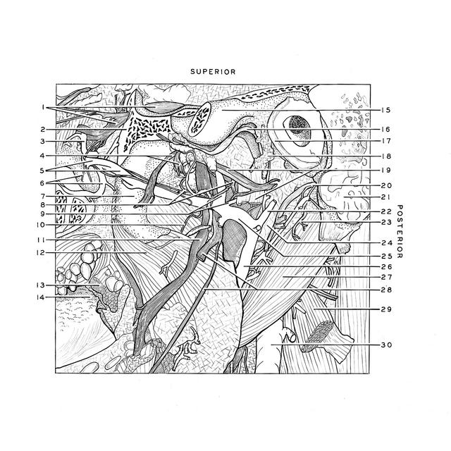
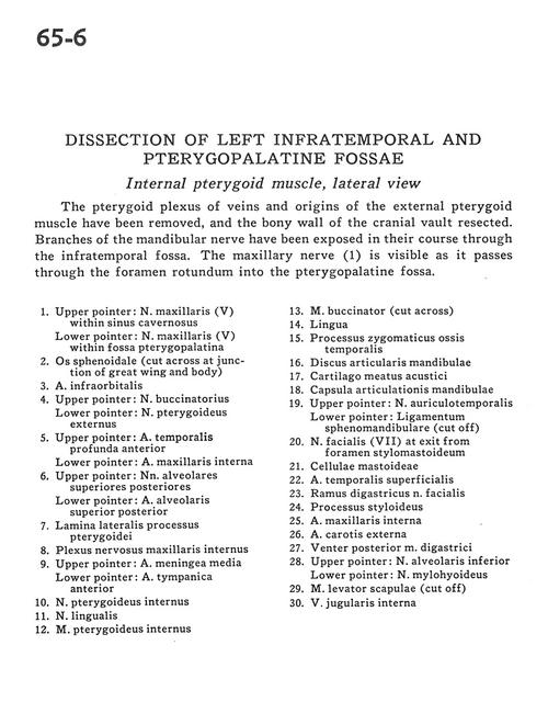
Dissection of left infratemporal and pterygopalatine fossae
Internal pterygoid muscle, lateral view
Stanford holds the copyright to the David L. Bassett anatomical images and has assigned
Creative Commons license Attribution-Share
Alike 4.0 International to all of the images.
For additional information regarding use and permissions,
please contact the Medical History Center.
Image #65-6
Dissection of left infratemporal and pterygopalatine fossae
Internal pterygoid muscle, lateral view
The pterygoid plexus of veins and origins of the external pterygoid muscle have been removed, and the bony wall of the cranial vault resected. Branches of the mandibular nerve have been exposed in their course through the infratemporal fossa. The maxillary nerve (1) is visible as it passes through the foramen rotundum into the pterygopalatine fossa.
- Upper pointer: Maxillary nerve (V) within cavernous sinus Lower pointer: Maxillary nerve (V) within Pterygopalatine fossa
- Sphenoid bone (cut across at junction of great wing and body)
- Infraorbital artery
- Upper pointer: Buccal nerve Lower pointer: External pterygoid nerve
- Upper pointer: Deep anterior temporal artery Lower pointer: Internal maxillary artery
- Upper pointer: Superior posterior alveolar nerves Lower pointer: Superior posterior alveolar artery
- Lateral plate of pterygoid process
- Internal maxillary nerve plexus
- Upper pointer: Middle meningeal artery Lower pointer: Anterior tympanic artery
- Internal pterygoid nerve
- Lingual nerve
- Internal pterygoid muscle
- Buccinator muscle (cut across)
- Tongue
- Zygomatic process of temporal bone
- Articular disc of mandible
- Cartilaginous acoustic meatus
- Joint capsule of mandible
- Upper pointer: Auriculotemporal nerve Lower pointer: Sphenomandibular ligament (cut off)
- Facial nerve (VII) at exit from stylomastoid foramen
- Mastoid cells
- Superficial temporal artery
- Digastric branch of facial nerve
- Styloid process
- Internal maxillary artery
- External carotid artery
- Posterior belly of digastric muscle
- Upper pointer: Inferior alveolar nerve Lower pointer: Mylohyoid nerve
- Levator scapulae muscle (cut off)
- Internal jugular vein