Dissection of left parotideomasseteric region
Distribution of nerves and blood vessels within masseter muscle, anterolateral view
Stanford holds the copyright to the David L. Bassett anatomical images and has assigned
Creative Commons license Attribution-Share
Alike 4.0 International to all of the images.
For additional information regarding use and permissions,
please contact the Medical History Center.
Image #63-6
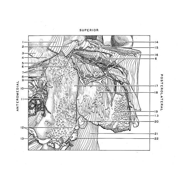
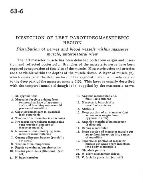
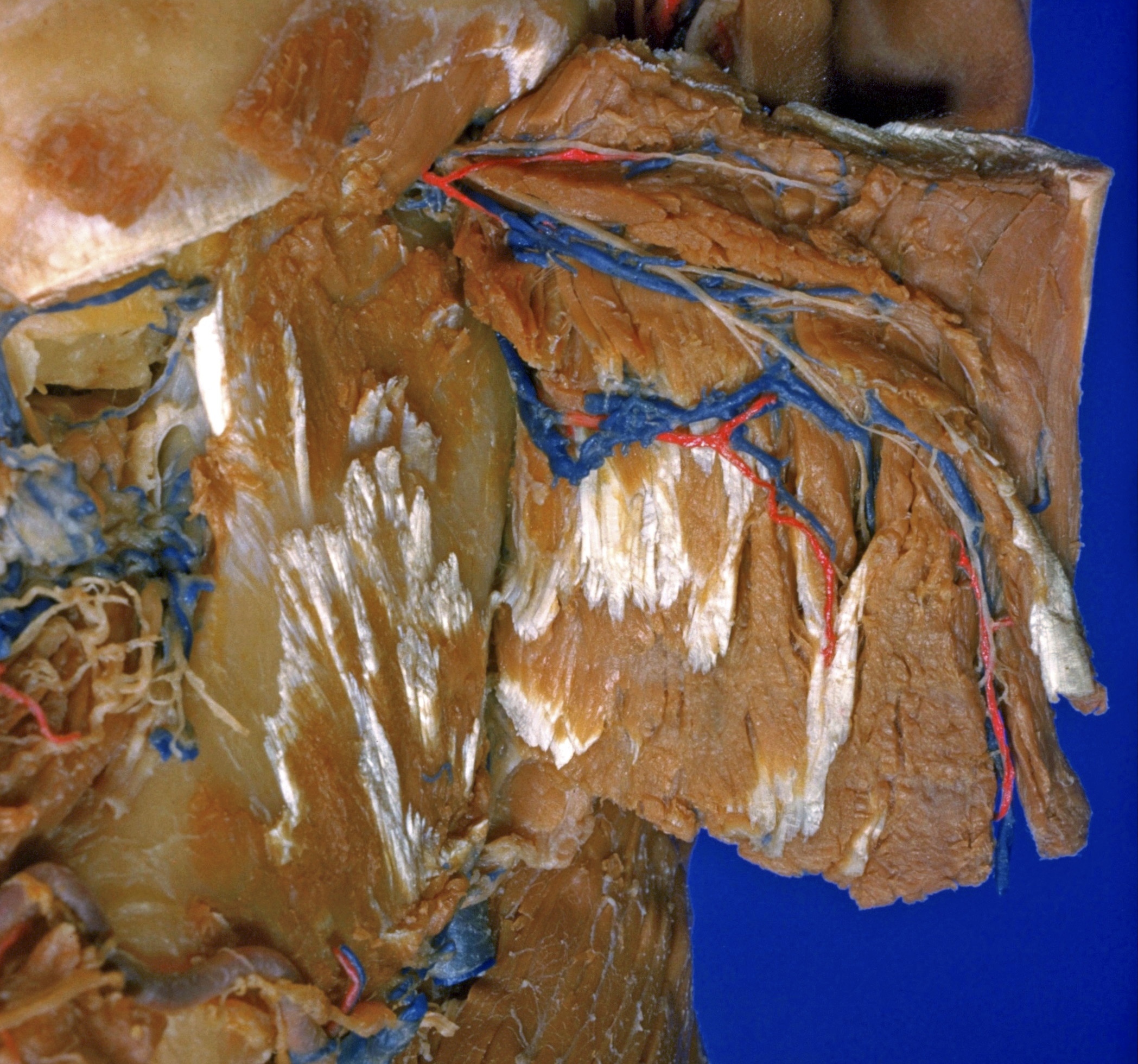
Dissection of left parotideomasseteric region
Distribution of nerves and blood vessels within masseter muscle, anterolateral view
The left masseter muscle has been detached both from the origin and insertion, and reflected posteriorly. Branches of the masseteric nerve have been exposed by separation of fascicles of the muscle. Masseteric veins, and arteries are also visible within the depths of the muscle tissue. A layer of muscle (2), which arises from the deep surface of the zygomatic arch, is closely related to the deep part of the masseter muscle (15). This layer is usually described with the temporal muscle although it is supplied by the masseteric nerve.
- Zygomatic muscle
- Muscular fascicle arising from temporal surface of zygomatic arch and inserting on coronoid process of mandible
- Zygomatic head of levator labii superioris muscle
- Tendon of masseter muscle (cut across)
- Coronoid process mandible (cut muscle fibers are of masseter muscle)
- Masseteric nerve (emerging from mandibular incisure)
- Buccal fat pad (partially cut away)
- Tendon of temporalis muscle
- Fascia covering buccinator nerve
- Parotid duct (cut off)
- Buccinator nerve
- Angle of mandible and external maxillary artery
- Masseteric branch of external maxillary artery
- Auricle
- Deep portion of masseter muscle (cut across near origin from zygomatic arch)
- Anterior margin of masseter muscle (reflected)
- Branch of mandible
- Deep portion of masseter muscle cut away from insertion into ramus of mandible
- Superficial portion of masseter muscle cut away from insertion into body of mandible
- Parotid gland
- Sternocleidomastoid muscle
- Posterior facial vein (cut off)