Dissection of ear from lateral aspect
Left tympanic membrane, anteroinferior view
Stanford holds the copyright to the David L. Bassett anatomical images and has assigned
Creative Commons license Attribution-Share
Alike 4.0 International to all of the images.
For additional information regarding use and permissions,
please contact the Medical History Center.
Image #60-3
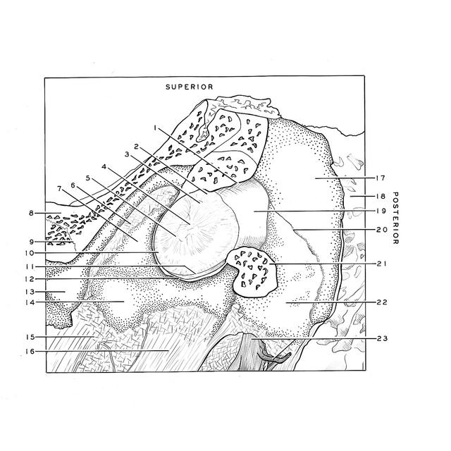
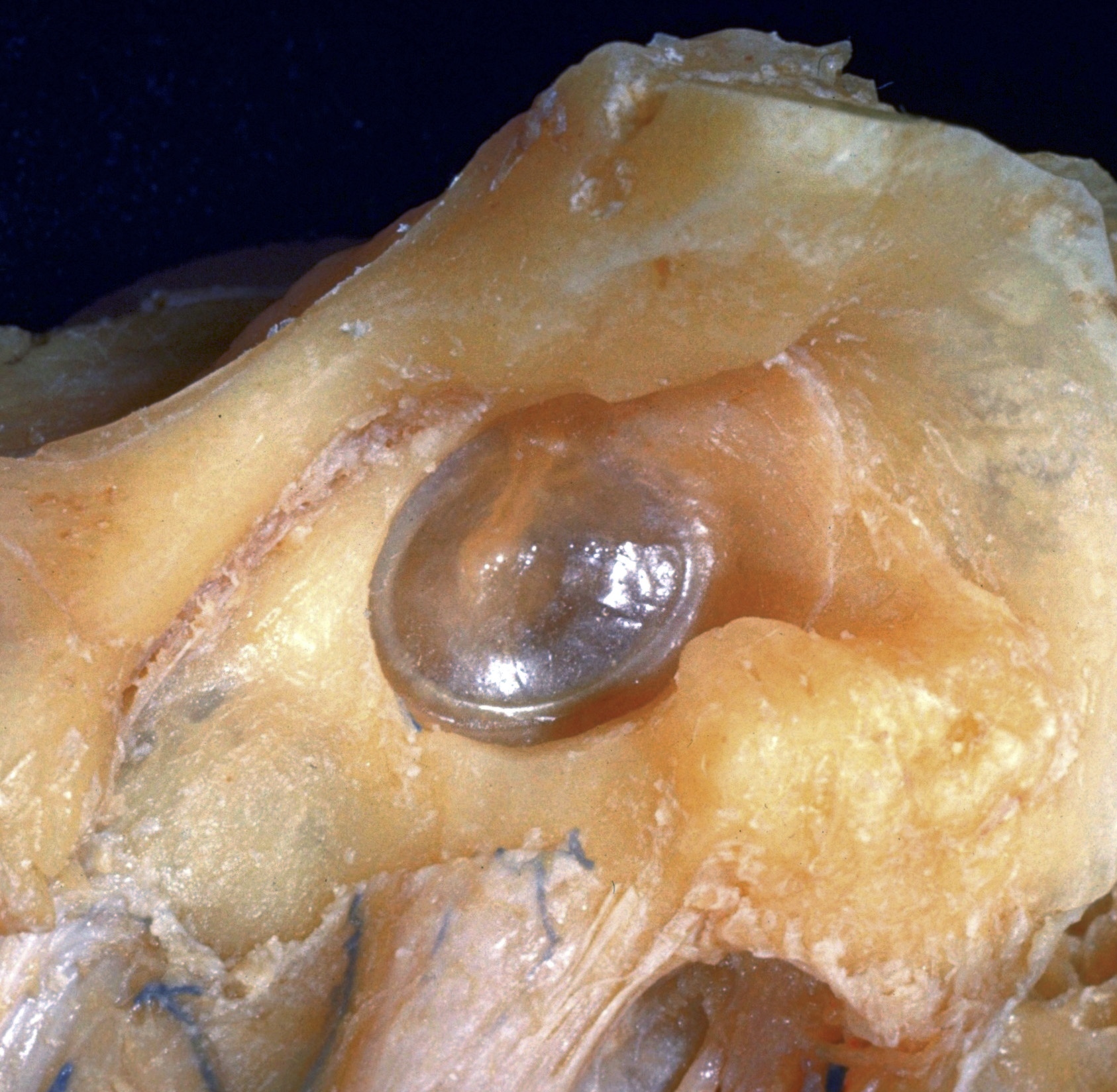
Dissection of ear from lateral aspect
Left tympanic membrane, anteroinferior view
The anterior wall of the external auditory canal has been removed to reveal the tympanic membrane through which the manubrium of the malleus is visible. Due to the direction of view, however, the manubrium does not present the anterosuperior inclination customarily seen in otoscopic examination.
- Cut edge of bony roof of external auditory canal
- Flaccid part of tympanic membrane
- Lateral process malleus (forming malleolar prominence)
- Manubrium of malleus (visible as malleolar stria)
- Pars tensa tympanic membrane
- Petrotympanic fissure
- External surface of tympanic annulus seen at 3 in previous view
- Temporal bone (squamous part)
- Sphenosquamous suture
- Limbus tympanic membrane
- Annulus fibrocartilaginous tympanic membrane
- Cut edge of osseous floor of auditory canal
- Angular spine
- Tympanic part temporal bone
- Alar fascia
- Styloid process
- Temporal bone (squamous part) forming roof of external acoustic meatus
- Mastoid cells
- Skin lining external acoustic meatus
- Tympanomastoid suture
- Cut edge of tympanic part of temporal bone
- Lateral margin of tympanic part of temporal bone
- Facial nerve (VII)