Exploration of the brain from its basal aspect
Tail of the caudate nucleus and retrolenticular part of the internal capsule
Stanford holds the copyright to the David L. Bassett anatomical images and has assigned
Creative Commons license Attribution-Share
Alike 4.0 International to all of the images.
For additional information regarding use and permissions,
please contact the Medical History Center.
Image #6-5
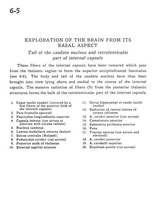
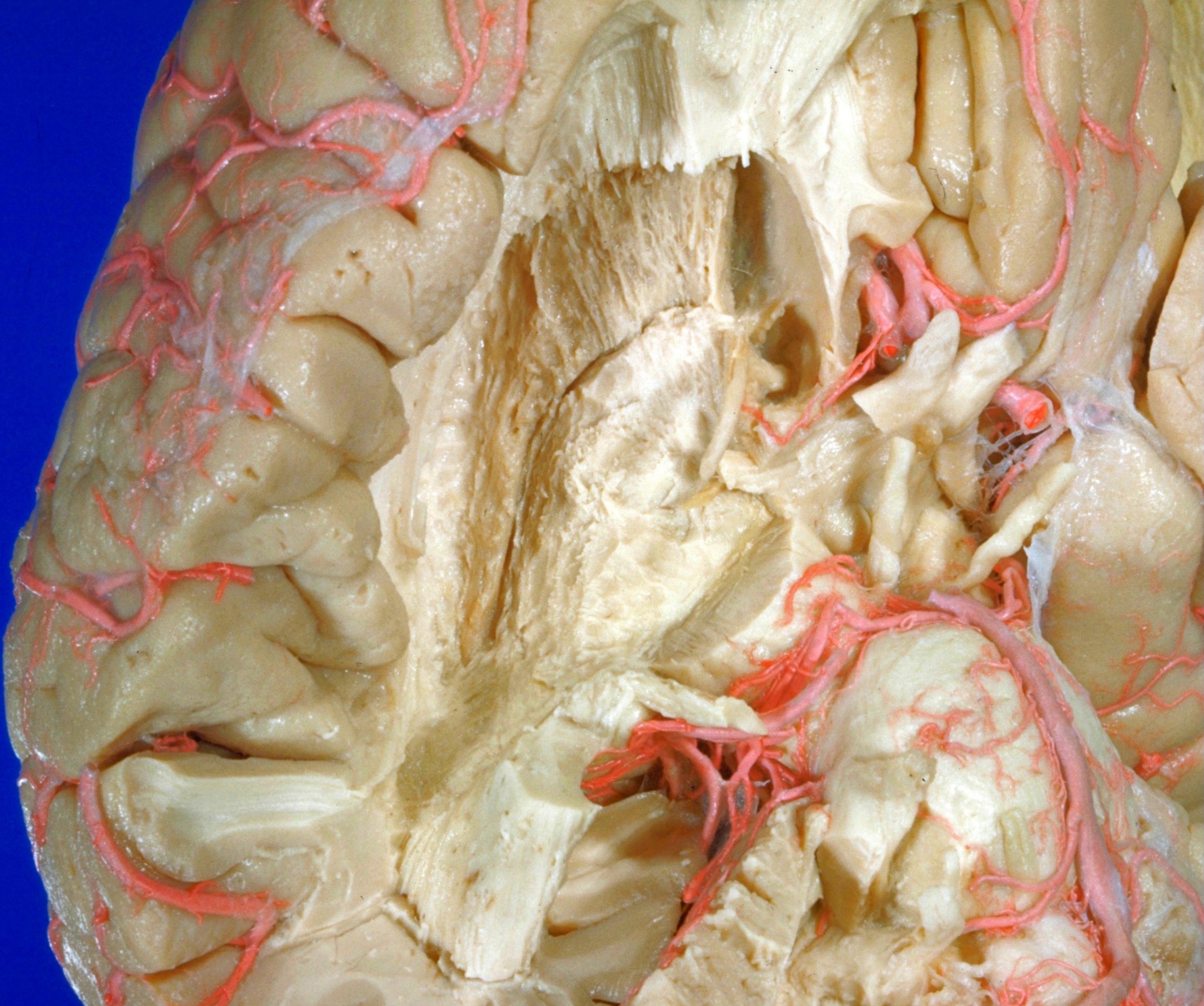
Exploration of the brain from its basal aspect
Tail of the caudate nucleus and retrolenticular part of the internal capsule
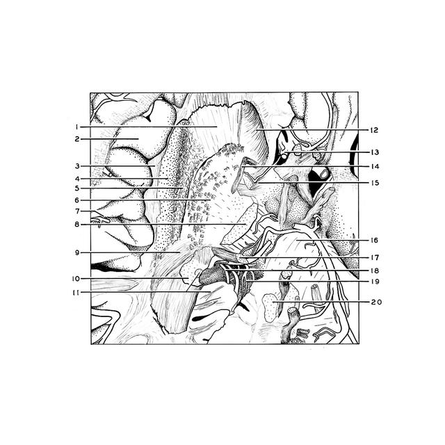
Those fibers of the internal capsule have been removed which pass from the thalamic region to form the superior occipitofrontal fasciculus (see 6-6). The body and tail of the caudate nucleus have thus been brought into view lying above and medial to the course of the internal capsule. The massive radiation of fibers (9) from the posterior thalamic structures forms the bulk of the retrolenticular part of the internal capsule.
- Head of caudate nucleus (covered by a few fibers of the anterior limb of the internal capsule)
- Frontal part of operculum
- Superior longitudinal fasciculus
- Internal capsule (cut across at junction with corona radiata)
- Caudate nucleus
- External medullary lamina (thalamus)
- Central sulcus (Rolandic)
- Cerebral peduncle (cut across)
- Posterior stalk of thalamus
- External sagittal stratum
- Hippocampal gyrus and caudate nucleus (tail)
- Radiation of rostral lamina of corpus callosum
- Anterior cerebral artery (cut across)
- Anterior commissure
- Anterior perforated substance
- Pons
- Optic tract (cut across and elevated)
- Posterior cerebral artery
- Superior cerebellar artery
- Brachium pontis (middle cerebellar peduncle) (cut across)