Exploration of the brain from its basal aspect
Internal capsule
Stanford holds the copyright to the David L. Bassett anatomical images and has assigned
Creative Commons license Attribution-Share
Alike 4.0 International to all of the images.
For additional information regarding use and permissions,
please contact the Medical History Center.
Image #6-1
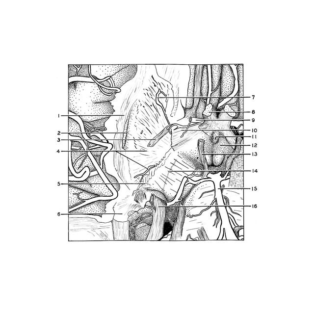
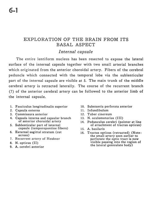
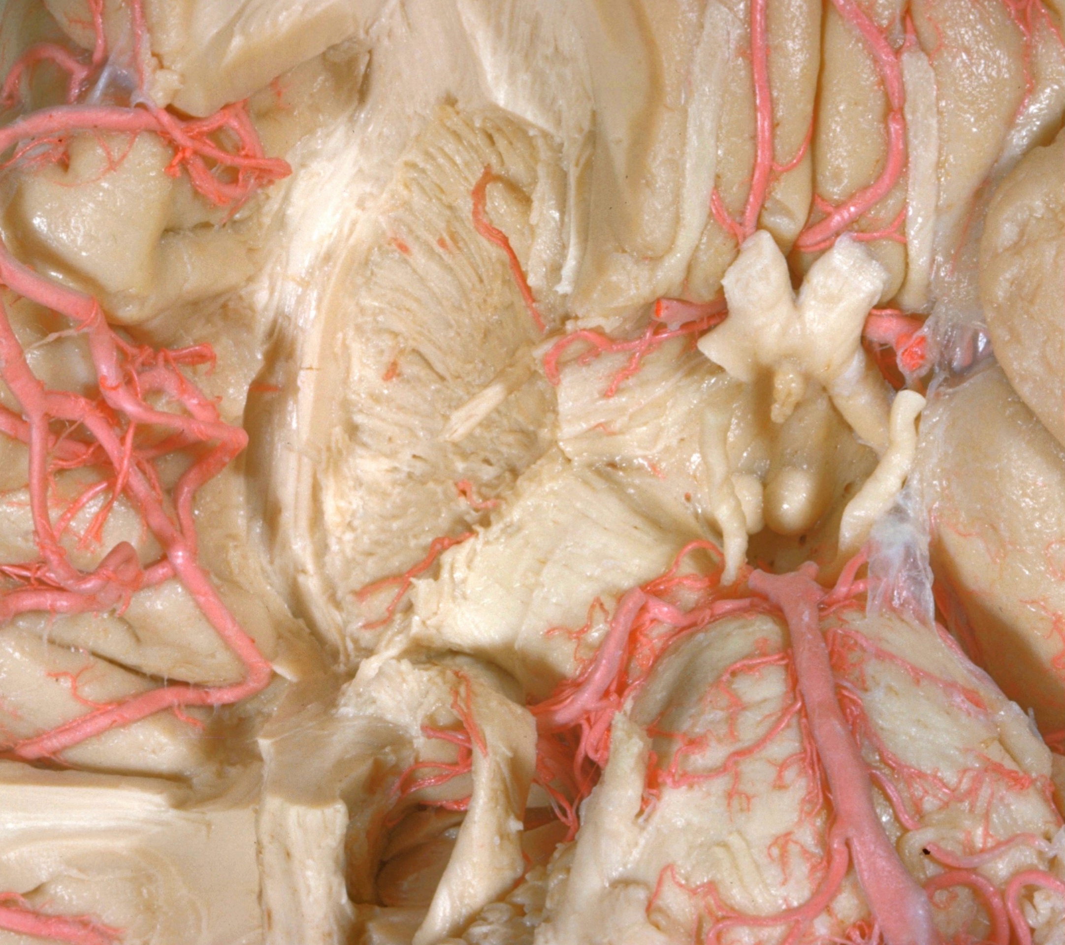
Exploration of the brain from its basal aspect
Internal capsule
The entire lentiform nucleus has been resected to expose the lateral surface of the internal capsule together with two small arterial branches which originated from the anterior choroidal artery. Fibers of the cerebral peduncle which connected with the temporal lobe via the sublenticular part of the internal capsule are visible at 5. The main trunk of the middle cerebral artery is retracted laterally. The course of the recurrent branch (7) of the anterior cerebral artery can be followed to the anterior limb of the internal capsule.
- Superior longitudinal fasciculus
- External capsule
- Anterior commissure
- Internal capsule and capsular branch of anterior choroidal artery
- Sublenticular part of internal capsule (temporopontine fibers)
- External sagittal stratum (cut across)
- Recurrent artery of Heubner
- Optic nerve (II)
- Anterior cerebral artery
- Anterior perforated substance
- Infundibulum
- Tuber cinereum
- Oculomotor nerve (III)
- Cerebral peduncle (pointer at line of attachment of optic tract)
- Basilar artery
- Optic tract (retracted) (Note: the small artery seen earlier to perforate the optic tract is now visible passing into the region of the lateral geniculate body)