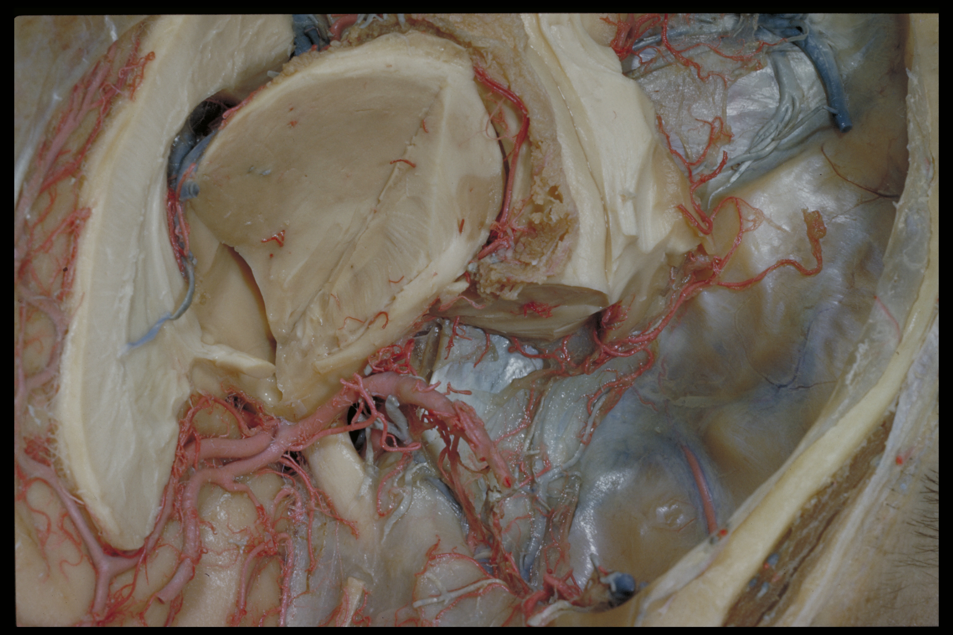
Microradiograph of eye; central optic pathways and related structures
Relations of optic pathways at base of brain.
Stanford holds the copyright to the David L. Bassett anatomical images and has assigned
Creative Commons license Attribution-Share
Alike 4.0 International to all of the images.
For additional information regarding use and permissions,
please contact the Medical History Center.
Image #58A-4
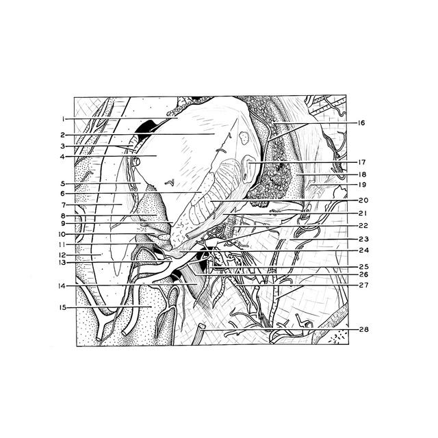
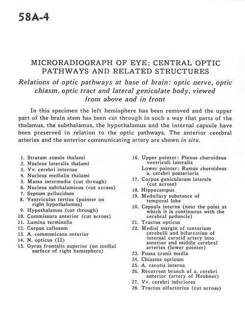
Microradiograph of eye; central optic pathways and related structures
Relations of optic pathways at base of brain.
In this specimen the left hemisphere has been removed and the upper part of the brain stem has been cut through in such a way that parts of the thalamus, the subthalamus, the hypothalamus and the internal capsule have been preserved in relation to the optic pathways. The anterior cerebral arteries and the anterior communicating artery are shown in situ.
- Stratum zonale thalami
- Lateral nucleus of thalamus
- Internal cerebral veins
- Medial nucleus of thalamus
- Massa intermedia (cut through)
- Subthalamic nucleus (cut across)
- Septum pellucidum
- Third ventricle (pointer on right hypothalamus)
- Hypothalamus (cut through)
- Anterior commissure (cut across)
- Lamina terminalis
- Corpus callosum
- Anterior communicating artery
- Optic nerve (II)
- Superior frontal gyrus (on medial surface of right hemisphere)
- Upper pointer: Lateral ventricular choroid plexus Lower pointer: Ramus choroideus ventriculi lateralis
- Lateral geniculate body
- Hippocampus
- Medullary substance of temporal lobe
- Internal capsule (near the point at which it is continuous with the cerebral peduncle)
- Optic tract
- Medial margin of the cerebral tentorium and bifurcation of internal carotid artery into anterior and middle cerebral arteries (lower pointer)
- Medical cranial fossa
- Optic chiasm
- Internal carotid artery
- Recurrent branch of anterior cerebral artery (artery of Heubner)
- Inferior cerebral veins
- Olfactory tract (cut across)
- [Legend above restored translation from Latin]