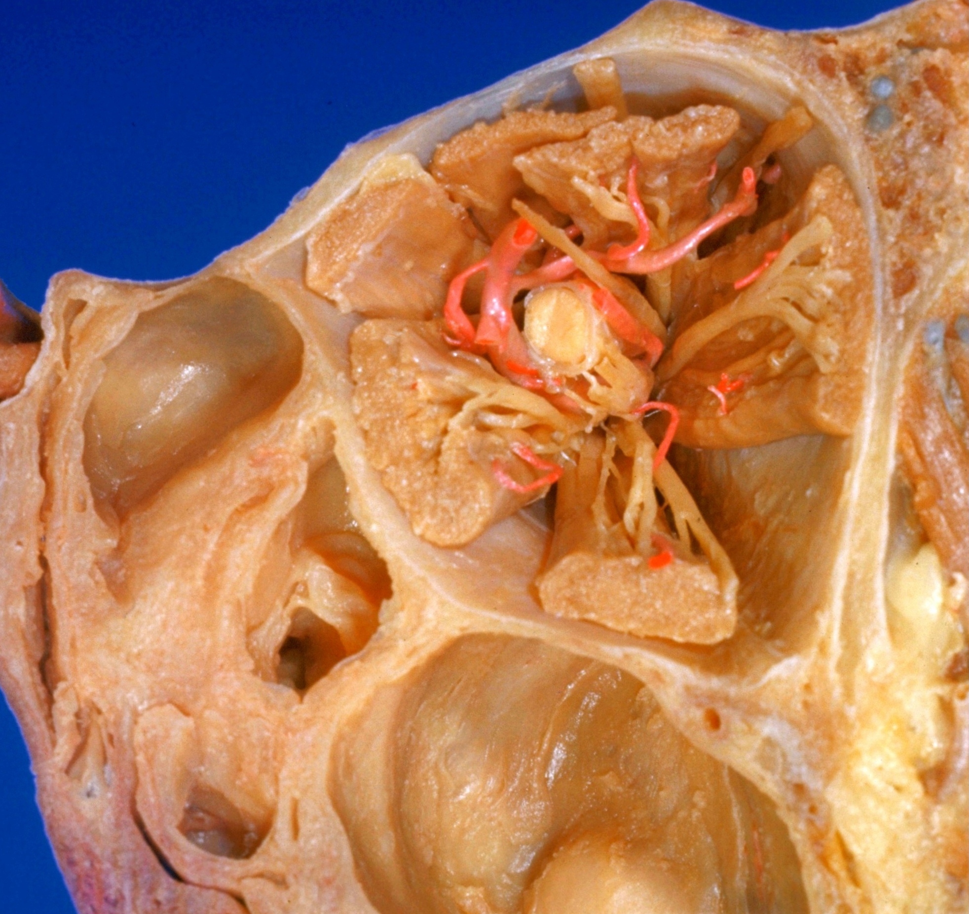
Apex of orbit
Relations of nerves, arteries and extraocular muscles, anterior view
Stanford holds the copyright to the David L. Bassett anatomical images and has assigned
Creative Commons license Attribution-Share
Alike 4.0 International to all of the images.
For additional information regarding use and permissions,
please contact the Medical History Center.
Image #57-3
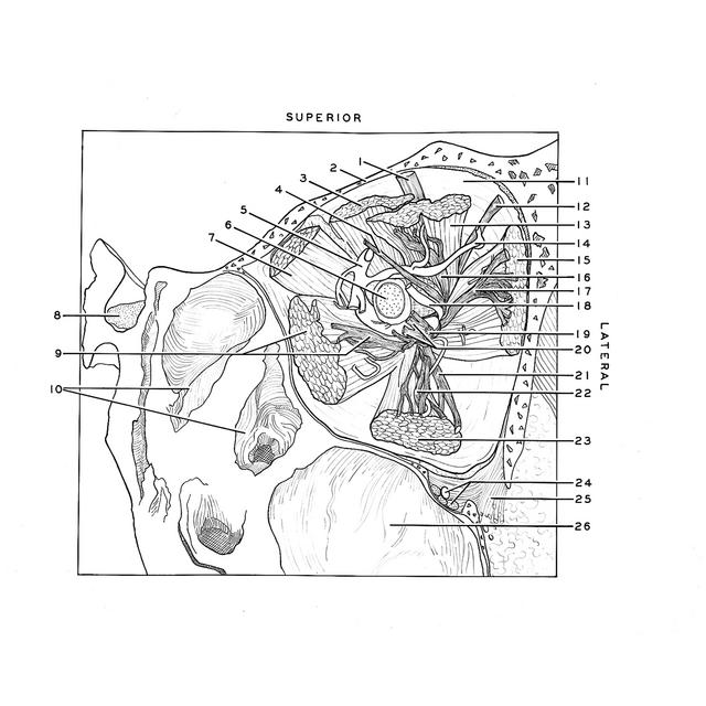
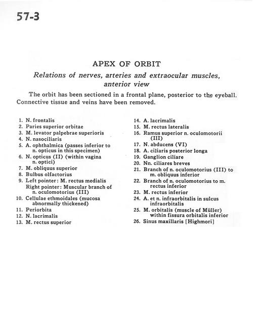
Apex of orbit
Relations of nerves, arteries and extraocular muscles, anterior view
The orbit has been sectioned in a frontal plane posterior to the eye. Fat, areolar connective tissue, nerves and blood vessels have been removed to demonstrate the fascia covering the extraordinary muscles and the eye. The fascia of the rectus muscles is thin posteriorly but becomes thick as the muscles approach the eye. It blends with the fascia of the bulb (Tenon's capsule) and, in addition, forms weblike folds (1) which extend between neighboring rectus muscles. The suspensory ligament of the eye has been described with the previous view.
- Frontal nerve
- Superior wall orbit
- Levator palpebrae superioris muscle
- Nasociliary nerve
- Ophthalmic artery (passes inferior to optic nerve in this specimen)
- Optic nerve (II) (within sheath of optic nerve)
- Superior oblique muscle
- Olfactory bulb
- Left pointer: Medial rectus muscle Right pointer: Muscular branch of oculomotor nerve (III)
- Ethmoidal cells (mucosa abnormally thickened)
- Periorbita
- Lacrimal nerve
- Superior rectus muscle
- Lacrimal artery
- Lateral rectus muscle
- Superior branch of oculomotor nerve (III)
- Abducens nerve (VI)
- Posterior ciliary artery (long)
- Ciliary ganglion
- Short ciliary nerves
- Branch of oculomotor nerve (III) to inferior oblique muscle
- Branch of oculomotor nerve to inferior rectus muscle
- Inferior rectus muscle
- Infraorbital artery and nerve in infraorbital sulcus
- Orbital muscle (muscle of Müller) within Inferior orbital fissure
- Maxillary sinus