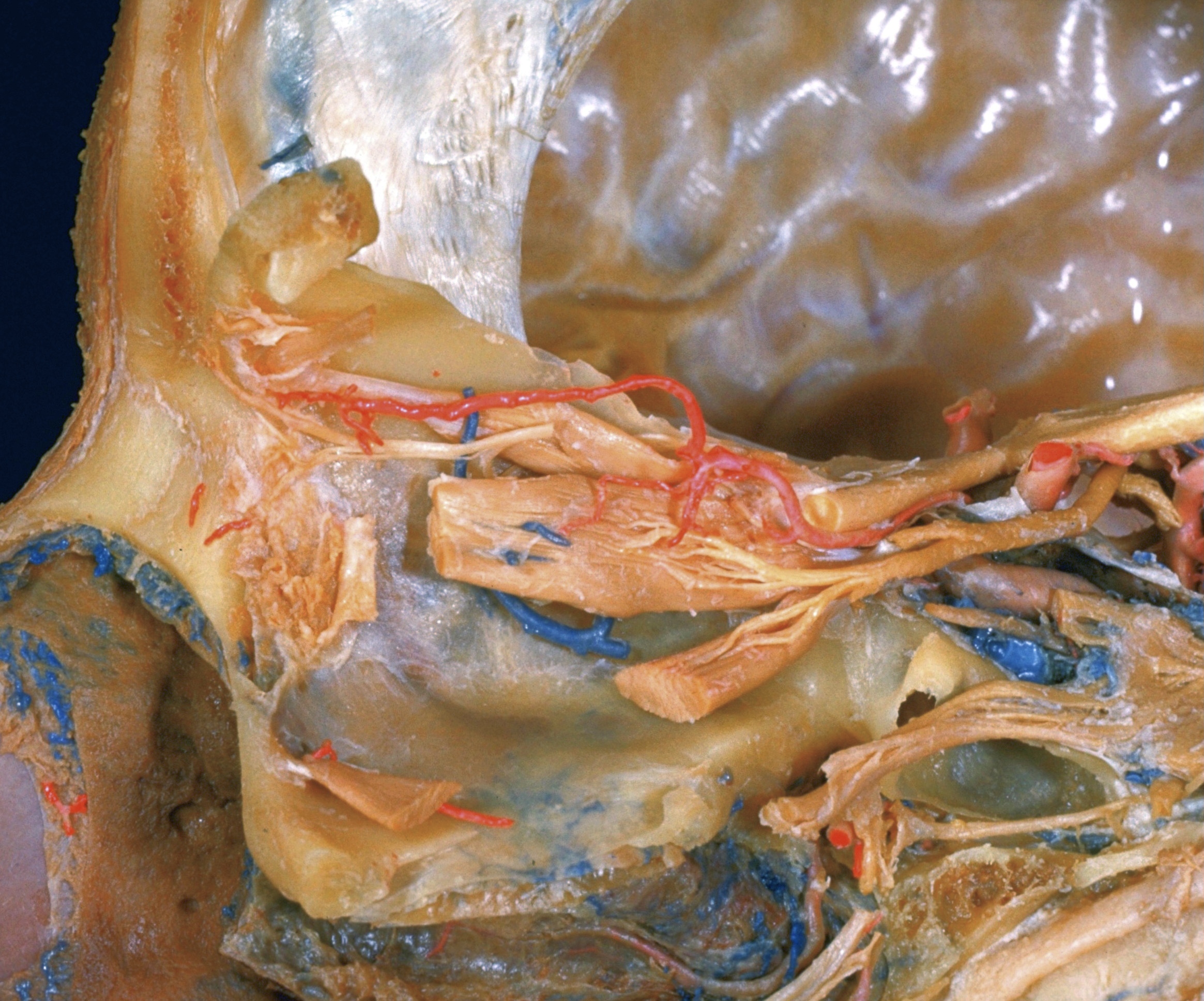
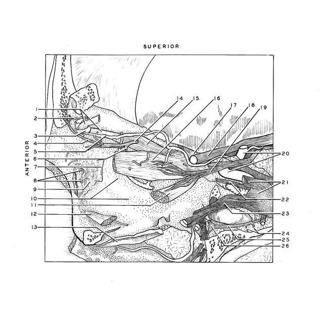
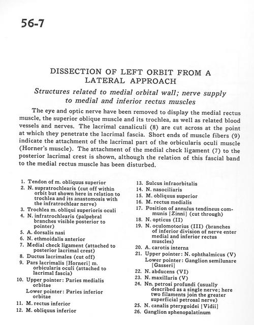
Dissection of left orbit from a lateral approach
Structures related to medial orbital wall; nerve supply to medial and inferior rectus muscles
Stanford holds the copyright to the David L. Bassett anatomical images and has assigned
Creative Commons license Attribution-Share
Alike 4.0 International to all of the images.
For additional information regarding use and permissions,
please contact the Medical History Center.
Image #56-7
Dissection of left orbit from a lateral approach
Structures related to medial orbital wall; nerve supply to medial and inferior rectus muscles
The eye and optic nerves have been removed to display the medial rectus muscle, the superior oblique muscle and its trochlea, as well as related blood vessels and nerves. The lacrimal canaliculi(8) are cut across at the point at which they penetrate the lacrimal fascia. Short ends of muscle fibres (9) indicate the attachment of the lacrimal part of the orbicularis oculi muscle (Horner's muscle). The attachment of the medial check ligament (7) to the posterior lacrimal crest is shown, although the relation of this fascial band to the medial rectus muscle has been disturbed.
- Tendon of superior oblique muscle
- Supratrochlear nerve (cut off within orbit but shown here in relation to trochlea and its anastomosis with the infratrochlear nerve)
- Superior oblique muscle
- Infratrochlear nerve (palpebral branches visible posterior to pointer)
- Dorsal nasal artery
- Anterior ethmoidal nerve
- Medial check ligament (attached to posterior lacrimal crest)
- Lacrimal duct (cut off)
- Lacrirnal part orbicularis oculi muscle (attached to lacrimal fascia)
- Upper pointer: Medial wall of orbit Lower pointer: Inferior wall of orbit
- Inferior rectus muscle
- Inferior oblique muscle
- Infraorbital sulcus
- Nasociliary nerve
- Superior oblique muscle
- Medial rectus muscle
- Position of common annular tendon (cut through)
- Optic nerve (II)
- Oculomotor nerve (III) (branches of inferior division of nerve enter medial and inferior rectus muscles)
- Internal carotid artery
- Upper pointer: Ophthalmic nerve (VI) Lower pointer: Semilunar ganglion (trigeminal)
- Abducens nerve (VI)
- Maxillary nerve (V)
- Deep petrosal nerves (usually described as a single nerve, here two filaments join the greater superficial petrosal nerve)
- Vidian nerve (of pterygoid canal)
- Sphenopalatine ganglion