Dissection of left orbit from a lateral approach
Relation of muscle fascia to Tenon's capsule; insertion of lateral rectus and inferior oblique muscles
Stanford holds the copyright to the David L. Bassett anatomical images and has assigned
Creative Commons license Attribution-Share
Alike 4.0 International to all of the images.
For additional information regarding use and permissions,
please contact the Medical History Center.
Image #56-2
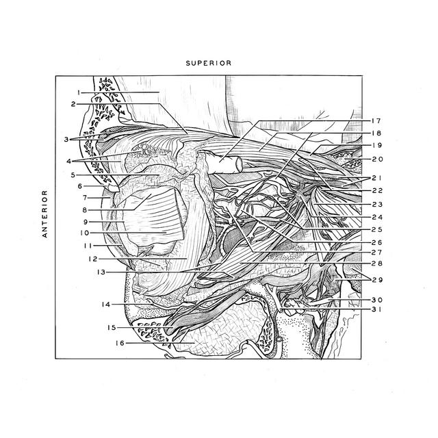
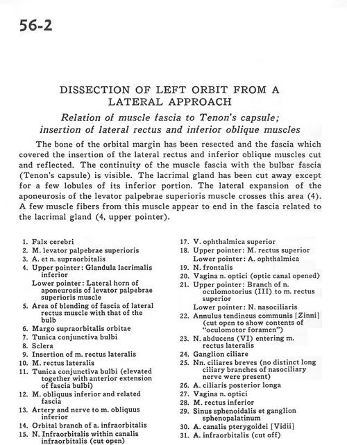
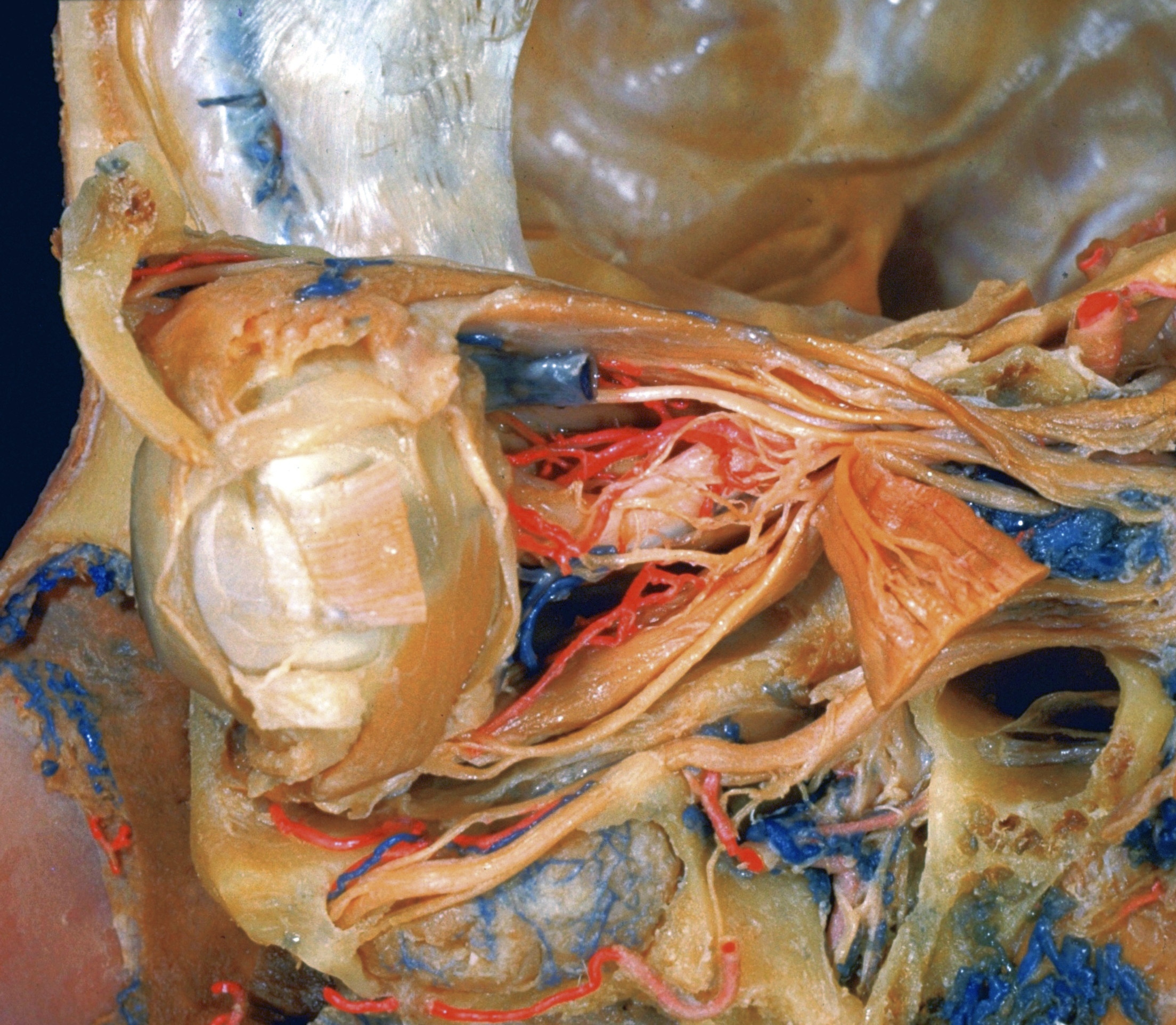
Dissection of left orbit from a lateral approach
Relation of muscle fascia to Tenon's capsule; insertion of lateral rectus and inferior oblique muscles
The bone of the orbital margin has been resected and the fascia which covered the insertion of the lateral rectus and inferior oblique muscles cut and reflected. The continuity of the muscle fascia with the bulbar fascia (Tenon's capsule) is visible. The lacrimal gland has been cut away except for a few lobules of its inferior portion. THe lateral expansion of the aponeurosis of the levator palpebrae superioris muscle crosses this area(4). A few muscle fibres from this muscle appear to end in a the fascia related to the lacrimal gland (4, upper pointer).
- Falx cerebri
- Levator palpebrae superioris muscle
- Supraorbital nerve and artery
- Upper pointer: Inferior lacrimal gland Lower pointer: Lateral horn of aponeurosis of levator palpebrae superioris muscle
- Area of blending of fascia of lateral rectus muscle with that of the bulh
- Supraorbital margin
- Tunica conjunctiva bulbar
- Sclera
- Insertion of lateral rectus muscle
- Lateral rectus muscle
- Tunica conjunctiva bulbar (elevated together with anterior extension of bulbar fascia)
- Inferior oblique muscle and related fascia
- Artery and nerve to inferior oblique muscle
- Orbital branch of infraorbital artery
- Infraorbital nerve within infraorbital canal (cut open)
- Upper pointer: Superior anterior alveolar nerve Lower pointer: Mucoperiosteum of maxillary sinus
- Superior ophthalmic vein
- Upper pointer: Superior rectus muscle Lower pointer: Ophthalmic artery
- Frontal nerve
- Sheath of optic nerve (optic canal opened)
- Upper pointer: Branch of oculomotor nerve (III) to superior rectus muscle Lower pointer: Nasociliary nerve
- Common annular tendon (cut open to show contents of "oculomotor foramen")
- Abducens nerve (VI) entering lateral rectus muscle
- Ciliary ganglion
- Short ciliary nerves (no distinct long ciliary branches of nasociliary nerve were present)
- Posterior ciliary artery (long)
- Sheath of optic nerve
- Inferior rectus muscle
- Sphenoid sinus and sphenopalatine ganglion
- Artery of pterygoid canal
- Infraorbital artery (cut off)