Dissection of left orbit from a lateral approach
Lateral rectus muscle reflected; ciliary nerves and ganglion
Stanford holds the copyright to the David L. Bassett anatomical images and has assigned
Creative Commons license Attribution-Share
Alike 4.0 International to all of the images.
For additional information regarding use and permissions,
please contact the Medical History Center.
Image #56-1
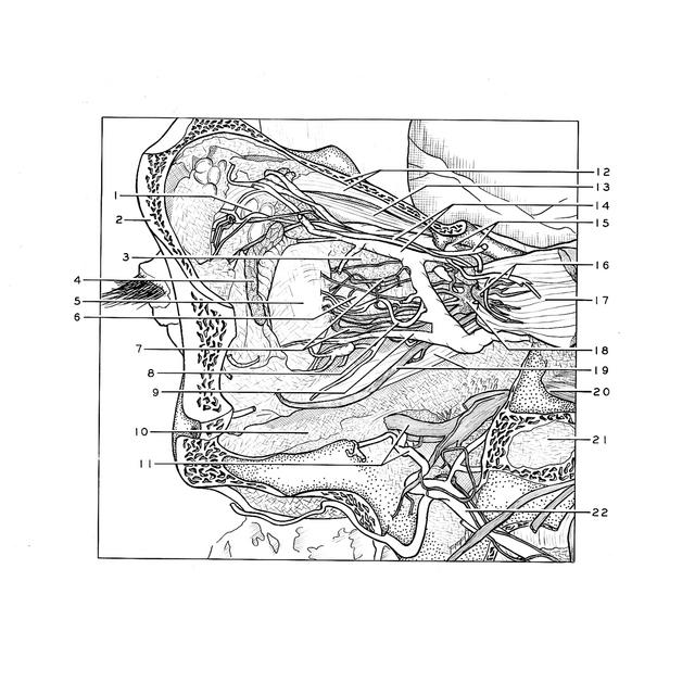
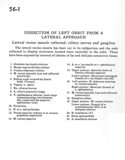
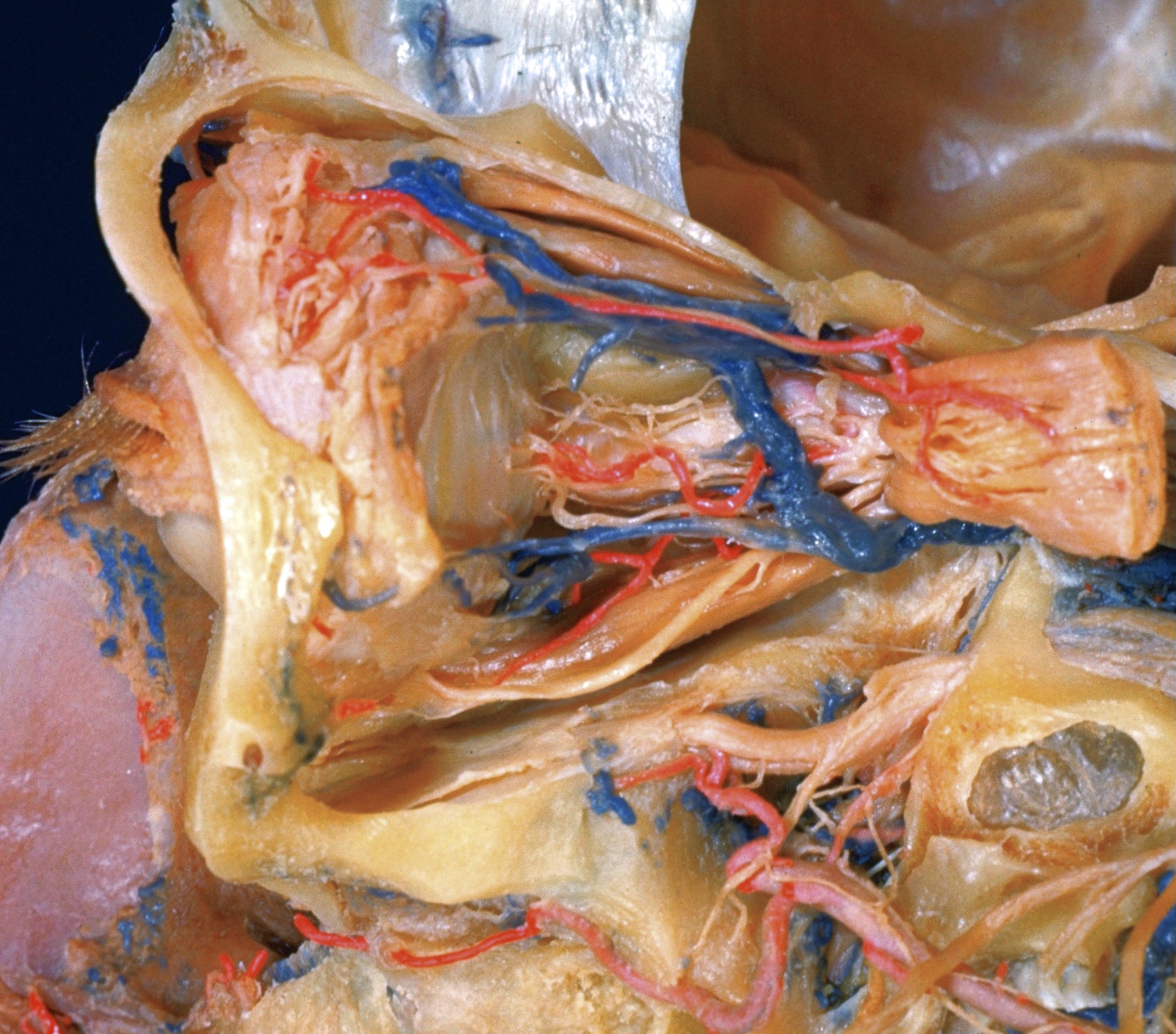
Dissection of left orbit from a lateral approach
Lateral rectus muscle reflected; ciliary nerves and ganglion
The lateral rectus muscle has been cut in its midportion and the ends reflected to display structures located more centrally in the orbit. These have been exposed by removal of lobules of fat and delicate connective tissue.
- Inferior lacrimal gland
- Supraorbital margin
- Orbital fat pad
- Lateral rectus muscle (cut end reflected anteriorly)
- Eyeball covered by bulbar fascia
- Sheath of optic nerve
- Short ciliary nerves
- Posterior ciliary artery (long)
- Inferior ophthalmic vein (note large communicating vein between this vessel and the superior ophthalmic vein)
- Periorbita
- Infraorbital artery and vein
- Superior wall orbit and levator palpebrae superioris muscle
- Superior rectus muscle
- Lacrimalis artery and vein and superior ophthalmic vein
- Upper pointer: Anterior limit of superior orbital fissure Lower pointer: Recurrent meningeal branch of lacrimal artery (cut off)
- Left pointer: Abducens nerve entering lateral rectus muscle Right pointer: Muscular branch of ophthalmic artery
- Lateral rectus muscle (cut and reflected posteriorly)
- Ciliary ganglion
- Upper pointer: Inferior rectus muscle Lower pointer: Branch of oculomotor nerve (III) to inferior oblique muscle
- Maxillary nerve (V)
- Sphenoid sinus
- Internal maxillary artery