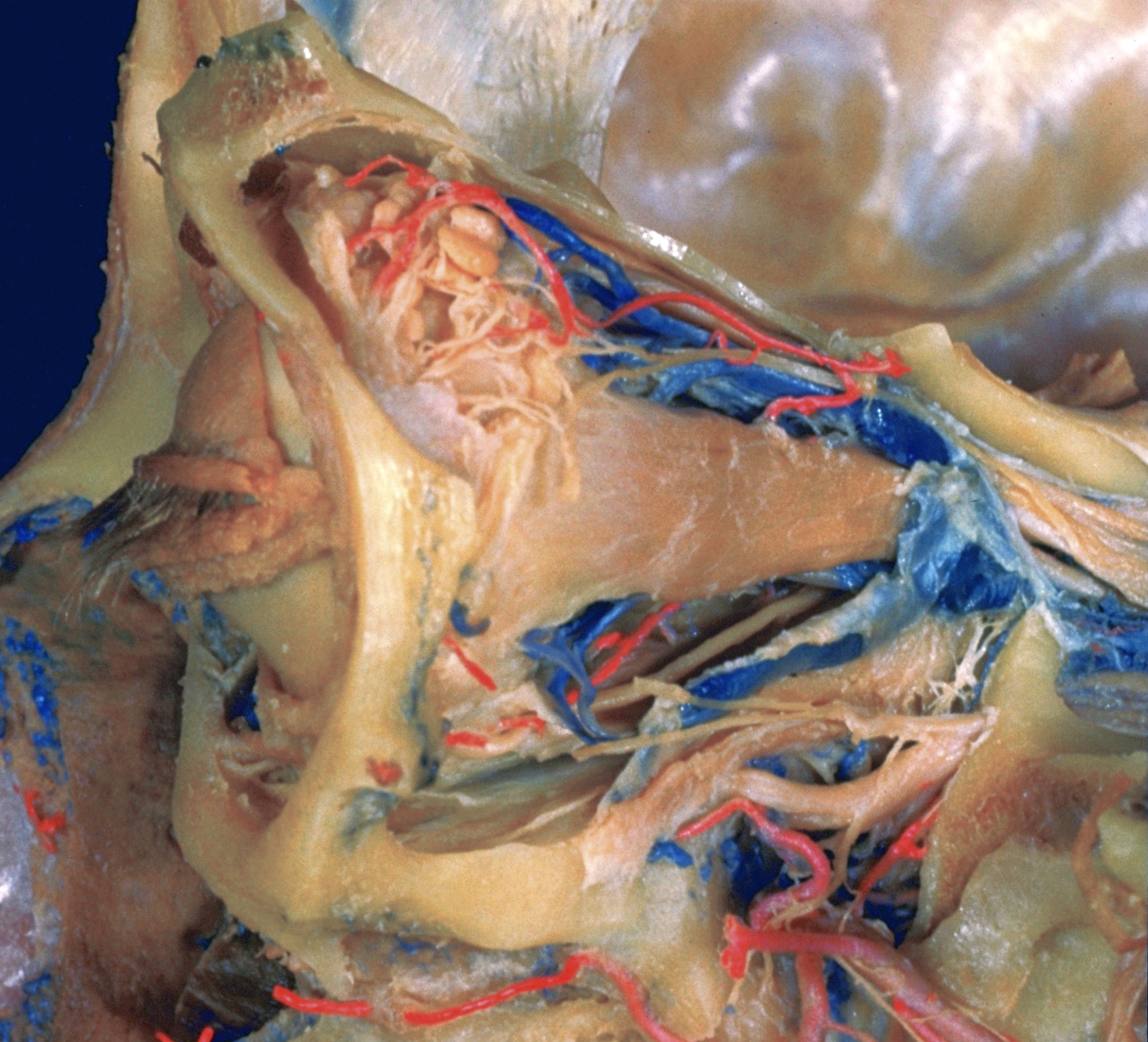
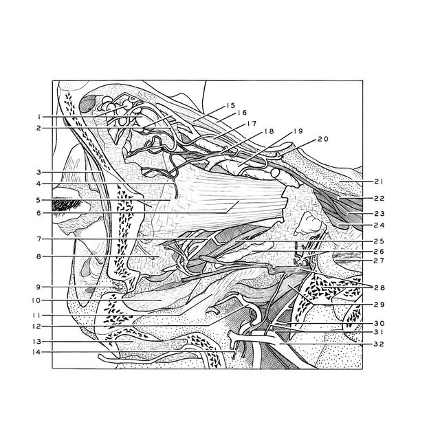
Dissection of left orbit from a lateral approach
Relation of orbital contents to structures within pterygopalatine fossa
Stanford holds the copyright to the David L. Bassett anatomical images and has assigned
Creative Commons license Attribution-Share
Alike 4.0 International to all of the images.
For additional information regarding use and permissions,
please contact the Medical History Center.
Image #55-6
Dissection of left orbit from a lateral approach
Relation of orbital contents to structures within pterygopalatine fossa
The lateral wall and part of the roof (16) of the orbit have been cut away and a narrow rim of bone left at the orbital margin (3) for orientation. The position of the superior and inferior orbital fissures may still be identified posteriorly near the roof and floor of the orbit because of the nerves (22,23,24), blood vessels (20,26), smooth muscle (11) and fascia which have been retained. For the size and shape of the fissures in the bony orbit reference should be made to 37-1.
- Remnant of superior lacrimal gland
- Lacrimal artery and vein
- Supraorbital margin
- Cut end of anastomotic branch of lacrimal nerve with zygomatic nerve
- Lateral check ligament
- Lateral rectus muscle
- Inferior musclar branch of ophthalmic artery
- Area of blending of fascias of lateral and inferior rectus muscles and inferior oblique muscle
- Zygomaticofacial branch of zygomatic nerve
- Periorbita of floor of orbit
- Orbital muscle (Müller's muscle situated within inferior orbital fissure)
- Infraorbital artery
- Mucoperiosteum of maxillary sinus
- Superior posterior alveolar nerve
- Levator palpebrae superioris muscle
- Superior wall orbit (cut away)
- Superior rectus muscle
- Lacrimal nerve
- Superior ophthalmic vein
- Recurrent branch of lacrimal artery which communicated through superior orbital fissure with middle meningeal artery)
- Anterior clinoid process
- Trochlear nerve (IV)
- Ophthalmic nerve (VI) entering Superior orbital fissure
- Abducens nerve (VI)
- Branch of oculomotor nerve (III) to inferior oblique muscle
- Upper pointer: Branch of Inferior ophthalmic vein Lower pointer: Orbital branch of sphenopalatine ganglion
- Foramen rotundum
- Upper pointer: Zygomatic nerve Lower pointer: Cut surface of greater wing of sphenoid bone
- Maxillary nerve (V2)
- Pharyngeal branch of artery of pterygoid canal
- Internal maxillary nerve plexus
- Internal maxillary artery