Dissection of left orbit from a lateral approach
Lateral rectus muscle; communication of lacrimal and zygomatic nerves
Stanford holds the copyright to the David L. Bassett anatomical images and has assigned
Creative Commons license Attribution-Share
Alike 4.0 International to all of the images.
For additional information regarding use and permissions,
please contact the Medical History Center.
Image #55-4
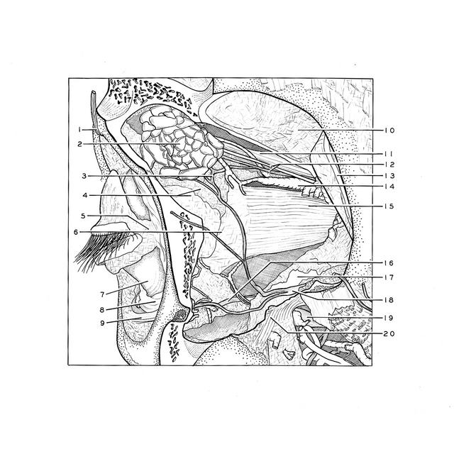
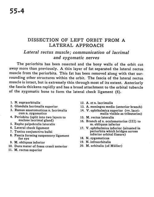
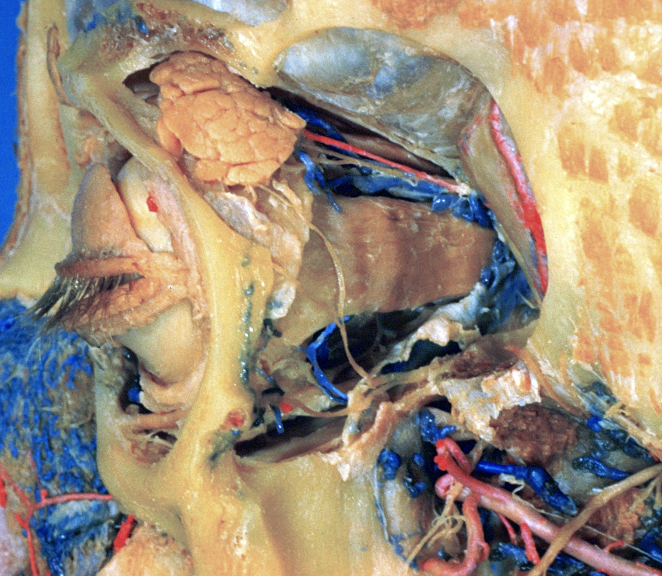
Dissection of left orbit from a lateral approach
Lateral rectus muscle; communication of lacrimal and zygomatic nerves
The periorbita has been resected and the bony walls of the orbit cut away more than previously. A thin layer of fat separated the lateral rectus muscle from the periorbita. This fat has been removed from along with that surrounding other structures within the orbit. The fascia of the lateral rectus muscle is intact, but is extremely thin through most of its extent. Anteriorly the fascia thickens rapidly and has a broad attachment to the orbital tubercle of the zygomatic bone to form the lateral check ligament (6).
- Supraorbital nerve
- Superior lacrimal gland
- Anastomotic branch of lacrimal nerve with zygomatic nerve
- Periorbita (split into two layers to enclose lacrimal gland)
- Lateral palpebral raphe
- Lateral check ligament
- Tunica conjunctiva
- Fascia forming suspensory ligament for eye
- Inferior oblique muscle
- Dura mater of anterior cranial fossa
- Superior rectus muscle
- Lacrimal artery and nerve
- Middle meningeal artery (anterior branch)
- Superior ophthalmic vein (lacrimal veins visible as tributaries)
- Lateral rectus muscle
- Branch of oculomotor nerve (III) to inferior oblique muscle
- Inferior ophthalmic vein (situated in periorbita which bridges across inferior orbital fissure)
- Zygomatic nerve
- Infraorbital nerve
- Orbital muscle (of Müller)