Dissection of right orbit from a superior approach
Ciliary ganglion and connections; nerve supply to medial, inferior and lateral rectus muscles
Stanford holds the copyright to the David L. Bassett anatomical images and has assigned
Creative Commons license Attribution-Share
Alike 4.0 International to all of the images.
For additional information regarding use and permissions,
please contact the Medical History Center.
Image #54-7
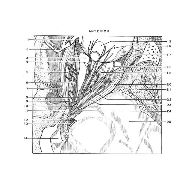
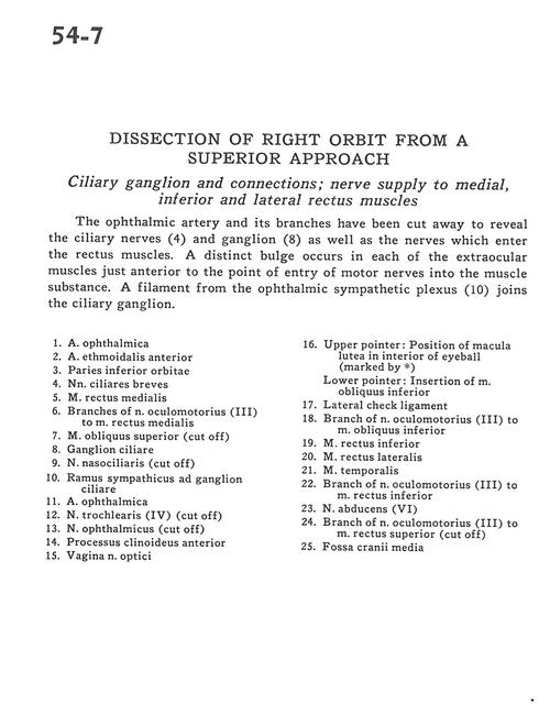
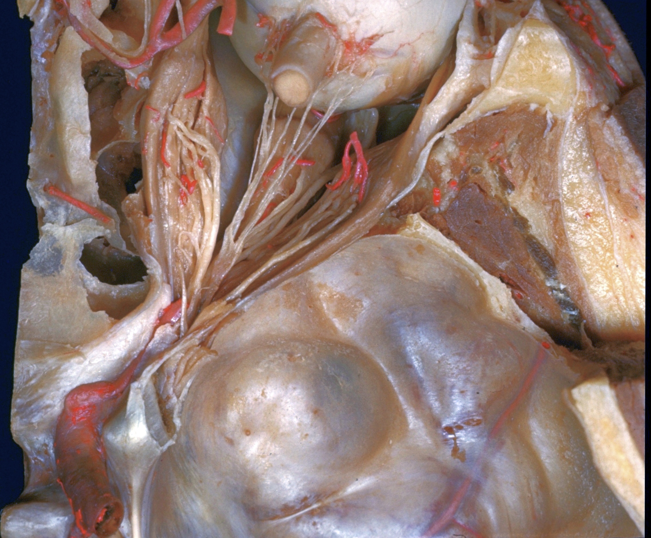
Dissection of right orbit from a superior approach
Ciliary ganglion and connections; nerve supply to medial, inferior and lateral rectus muscles
The ophthalmic artery and its branches have been cut away to reveal the ciliary nerves (4) and ganglion (8) as well as the nerves which enter the rectus muscles. A distinct bulge occurs in each of the extraocular muscles just anterior to the point of entry of motor nerves into the muscle substance. A filament from the ophthalmic sympathetic plexus (10) joins the ciliary ganglion.
- Ophthalmic artery
- Anterior ethmoidal artery
- Inferior wall of orbit
- Short ciliary nerves
- Medial rectus muscle
- Branches of oculomotor nerve (III) to medial rectus muscle
- Superior oblique muscle (cut off)
- Ciliary ganglion
- Nasociliary nerve (cut off)
- Filament from ophthalmic nerve plexus to ciliary ganglion
- Ophthalmic artery
- Trochlear nerve (IV) (cut off)
- Ophthalmic nerve (cut off)
- Anterior clinoid process
- Sheath of optic nerve
- Upper pointer: Position of macula lutea in interior of eyeball (marked by 4) Lower pointer: Insertion of inferior oblique muscle
- Lateral check ligament
- Branch of oculomotor nerve (III) to inferior oblique muscle
- Inferior rectus muscle
- Lateral rectus muscle
- Temporalis muscle
- Branch of oculomotor nerve (III) to inferior rectus muscle
- Abducens nerve (VI)
- Branch of oculomotor nerve (III) to superior rectus muscle (cut off)
- Middle cranial fossa