
Dissection of right orbit from a superior approach
Ophthalmic artery; ciliary nerves and arteries; sheath of optic nerve; insertion of superior oblique muscle
Stanford holds the copyright to the David L. Bassett anatomical images and has assigned
Creative Commons license Attribution-Share
Alike 4.0 International to all of the images.
For additional information regarding use and permissions,
please contact the Medical History Center.
Image #54-5
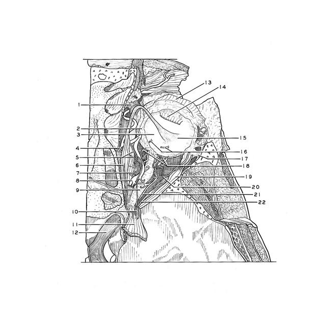
Dissection of right orbit from a superior approach
Ophthalmic artery; ciliary nerves and arteries; sheath of optic nerve; insertion of superior oblique muscle
The superior rectus muscle has been cut and reflected to expose underlying structures. The fascia of the bulb (Tenon's capsule) has been partially cut away. Short ends of two vorticose veins (16) protrude from the sclera. In this specimen the ophthalmic artery passes beneath the optic nerve rather than above it. This is a rather common variation.
- Superior oblique muscle
- Bulbar fascia
- Sclera
- Anterior ethmoidal artery
- Upper pointer: Infratrochlear nerve Lower pointer: Anterior ethmoidal nerve
- Superior oblique muscle
- Medial rectus muscle
- Ophthalmic artery
- Sheath of optic nerve
- Branch of oculomotor nerve entering superior rectus muscle
- Superior rectus muscle (cut and reflected)
- Optic nerve (II)
- Aponeurosis of levator palpebrae superioris muscle
- Superior rectus muscle (pointer near junction of muscle with tendon of insertion)
- Insertion of superior oblique muscle
- Vorticose veins (cut off)
- Lateral check ligament
- Short ciliary nerves
- Lateral rectus muscle
- Lacrimal artery
- Posterior ciliary artery
- Abducens nerve (VI)