Dissection of right orbit from a superior approach
Superior rectus muscle and related fascia
Stanford holds the copyright to the David L. Bassett anatomical images and has assigned
Creative Commons license Attribution-Share
Alike 4.0 International to all of the images.
For additional information regarding use and permissions,
please contact the Medical History Center.
Image #54-4
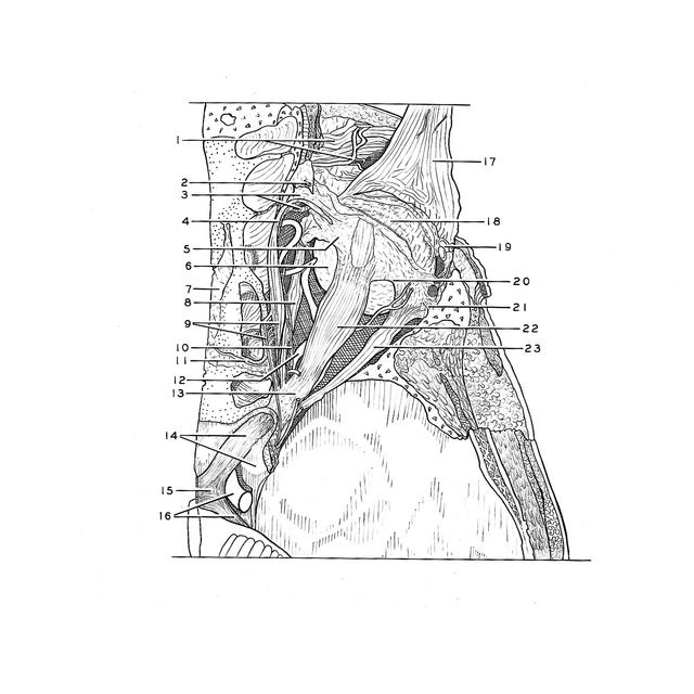
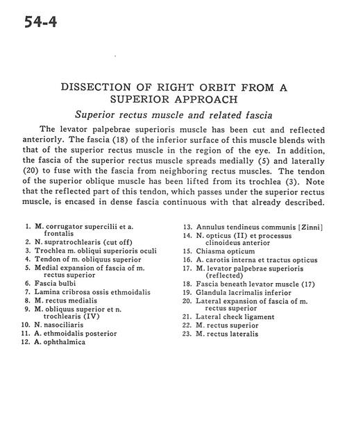
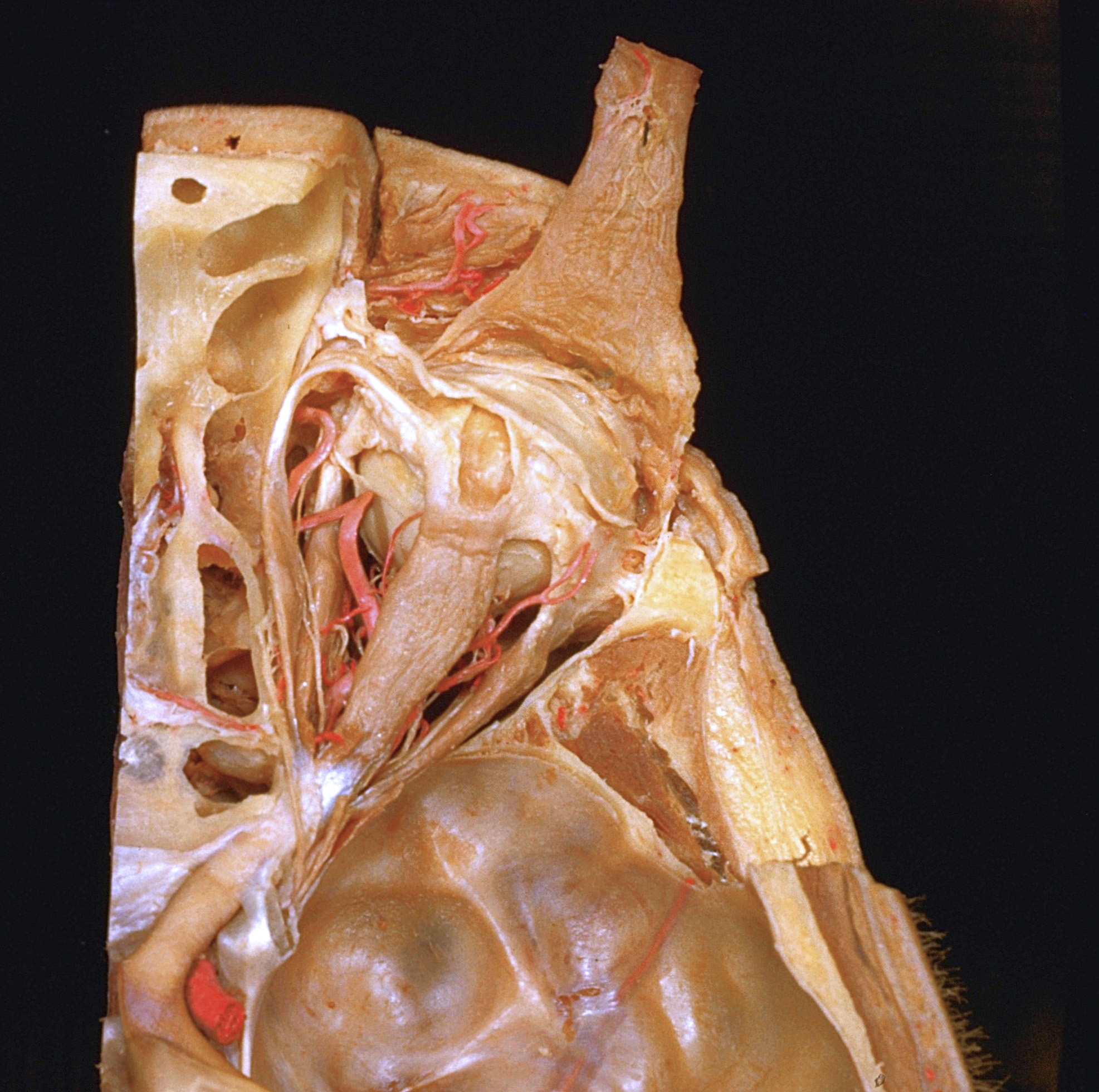
Dissection of right orbit from a superior approach
Superior rectus muscle and related fascia
The levator palpebrae superioris muscle has been cut and reflected anteriorly. The fascia (18) of the inferior surface of this muscle blends with that of the superior rectus muscle in the region of the eye. In addition, the fascia of the superior rectus muscle spreads medially (5) and laterally (20) to fuse with the fascia from neighboring rectus muscles. The tendon of the superior oblique muscle has been lifted from its trochlea (3). Note that the reflected part of this tendon, which passes under the superior rectus muscle, is encased in dense fascia continuous with that already described.
- Corrugator supercilii muscle and frontal artery
- Supratrochlear nerve (cut off)
- Superior oblique muscle
- Tendon of superior oblique muscle
- Medial expansion of fascia of superior rectus muscle
- Bulbar fascia
- Cribriform plate ethmoid bone
- Medial rectus muscle
- Superior oblique muscle and trochlear nerve (IV)
- Nasociliary nerve
- Artery ethmoidalis posterior
- Ophthalmic artery
- Common annular tendon
- Optic nerve (II) and anterior clinoid process
- Optic chiasm
- Internal carotid artery and optic tract
- Levator palpebrae superioris muscle (reflected)
- Fascia beneath levator muscle (17)
- Lacrimal gland (inferior)
- Lateral expansion of fascia of superior rectus muscle
- Lateral check ligament
- Superior rectus muscle
- Lateral rectus muscle