Dissection of left orbit from an anterior approach
Lacrimal sac and nasolacrimal duct opened
Stanford holds the copyright to the David L. Bassett anatomical images and has assigned
Creative Commons license Attribution-Share
Alike 4.0 International to all of the images.
For additional information regarding use and permissions,
please contact the Medical History Center.
Image #53-6
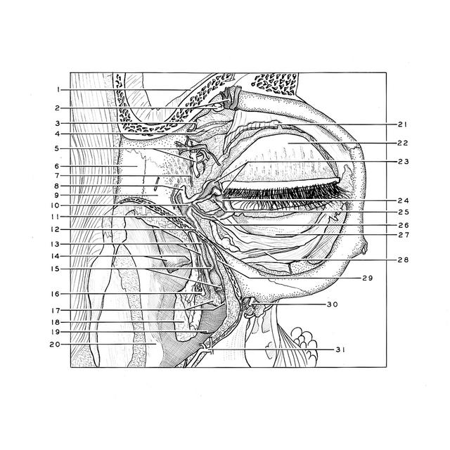
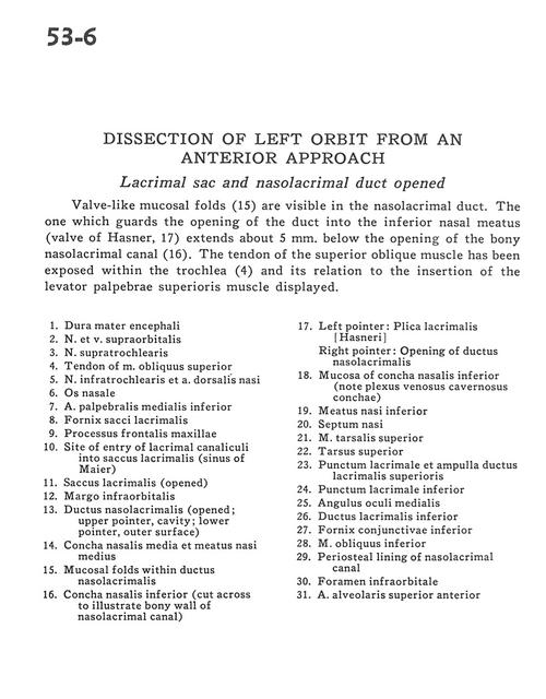
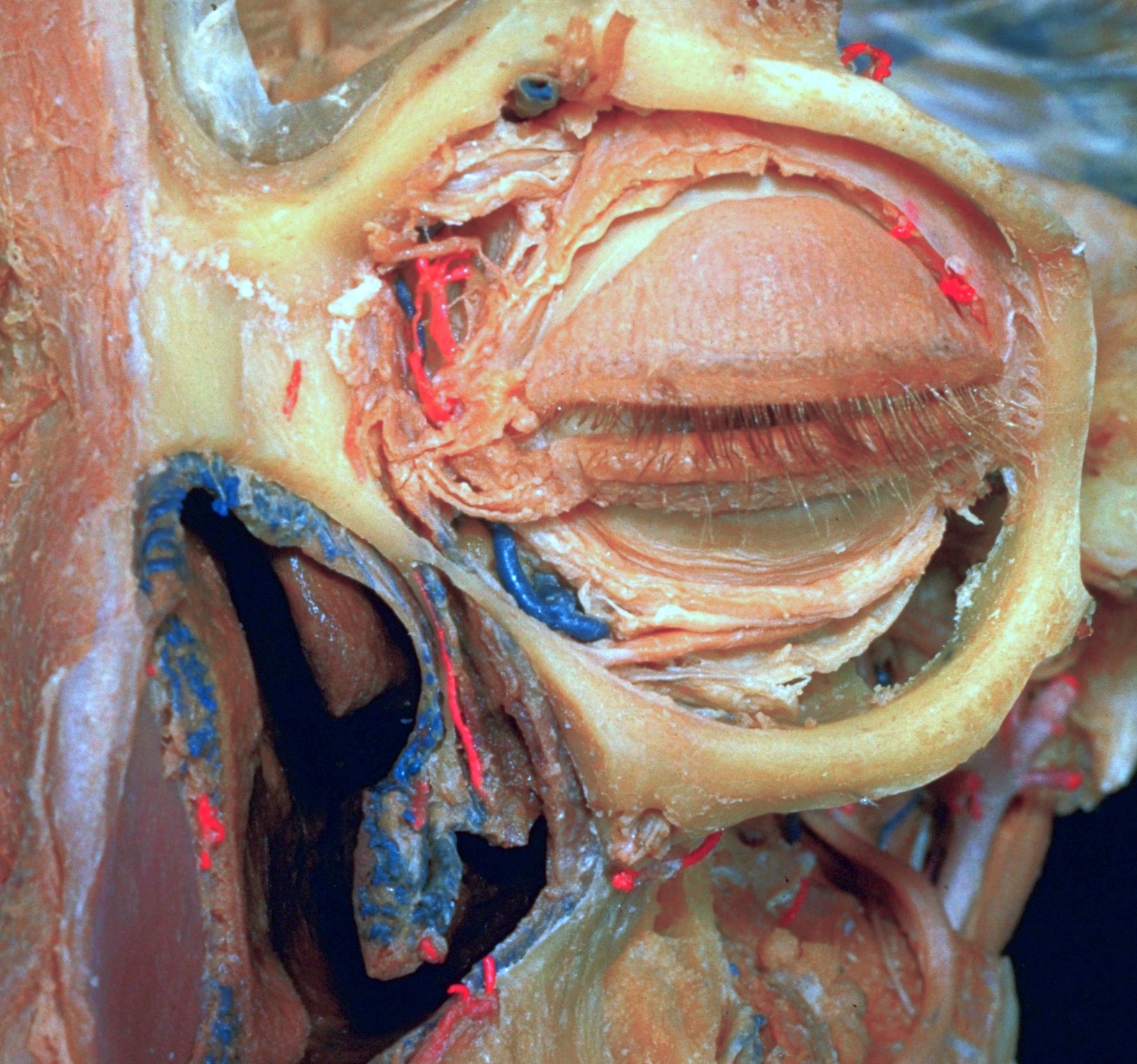
Dissection of left orbit from an anterior approach
Lacrimal sac and nasolacrimal duct opened
Valve-like mucosal folds (15) are visible in the nasolacrimal duct. The one which guards the opening of the duct into the inferior nasal meatus (valve of Hasner, 17) extends about 5 mm. below the opening of the bony nasolacrimal canal (16). The tendon of the superior oblique muscle has been exposed within the trochlea (4) and its relation to the insertion of the levator palpebrae superioris muscle displayed.
- Dura mater
- Supraorbital nerve and vein
- Supratrochlear nerve
- Tendon of superior oblique muscle
- Infratrochlear nerve and dorsal nasal artery
- Nasal bone
- Middle palpebral artery (inferior)
- Fornix lacrimal sac
- Frontal process of maxilla
- Site of entry of lacrimal canaliculi into lacrimal sac (sinus of Maier)
- Lacrimal sac (opened)
- Infraorbital margin
- Nasolacrimal duct (opened; upper pointer, cavity; lower pointer, outer surface)
- Middle nasal concha and middle nasal meatus
- Mucosal folds within nasolacrimal duct
- Inferior nasal concha (cut across to illustrate bony wall of nasolacrimal canal)
- Left pointer: Lacrimal folds Right pointer: Opening of nasolacrimal duct
- Mucosa of Inferior nasal concha (note venous plexus)
- Inferior nasal meatus
- Nasal septum
- Superior tarsalis muscle
- Superior tarsus
- Lacrimal puncta and ampulla of superior lacrimal duct
- Inferior Iacrimal punctum
- Medial ocular angle
- Inferior lacrimal duct
- Inferior conjunctival fornix
- Inferior oblique muscle
- Periosteal lining of nasolacrirnal canal
- Infraorbital foramen
- Superior anterior alveolar artery